 Movie
Movie Controller

[English] 日本語
 Yorodumi
- PDB-2f1g: Cathepsin S in complex with non-covalent 2-(Benzoxazol-2-ylamino)... -
+ Open data

- Basic information
| Entry | Database: PDB / ID: 2f1g | ||||||
|---|---|---|---|---|---|---|---|
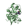
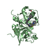
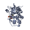
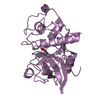
| Title | Cathepsin S in complex with non-covalent 2-(Benzoxazol-2-ylamino)-acetamide | ||||||
 Components Components | Cathepsin S | ||||||
 Keywords Keywords | HYDROLASE / Cathepsin S / noncovalent / inhibition / 2-(Benzooxazol-2-ylamino) acetamides | ||||||
| Function / homology |  Function and homology informationcathepsin S / basement membrane disassembly / positive regulation of cation channel activity / antigen processing and presentation of peptide antigen / endolysosome lumen / response to acidic pH / cellular response to thyroid hormone stimulus / Trafficking and processing of endosomal TLR / proteoglycan binding / Assembly of collagen fibrils and other multimeric structures ...cathepsin S / basement membrane disassembly / positive regulation of cation channel activity / antigen processing and presentation of peptide antigen / endolysosome lumen / response to acidic pH / cellular response to thyroid hormone stimulus / Trafficking and processing of endosomal TLR / proteoglycan binding / Assembly of collagen fibrils and other multimeric structures / toll-like receptor signaling pathway / cysteine-type endopeptidase activator activity involved in apoptotic process / fibronectin binding / antigen processing and presentation / collagen catabolic process / extracellular matrix disassembly / laminin binding / phagocytic vesicle / positive regulation of apoptotic signaling pathway / collagen binding / MHC class II antigen presentation / Degradation of the extracellular matrix / proteolysis involved in protein catabolic process / lysosomal lumen / Endosomal/Vacuolar pathway / protein processing / antigen processing and presentation of exogenous peptide antigen via MHC class II / late endosome / tertiary granule lumen / collagen-containing extracellular matrix / ficolin-1-rich granule lumen / adaptive immune response / lysosome / immune response / cysteine-type endopeptidase activity / serine-type endopeptidase activity / intracellular membrane-bounded organelle / Neutrophil degranulation / proteolysis / extracellular space / extracellular region Function and homology informationcathepsin S / basement membrane disassembly / positive regulation of cation channel activity / antigen processing and presentation of peptide antigen / endolysosome lumen / response to acidic pH / cellular response to thyroid hormone stimulus / Trafficking and processing of endosomal TLR / proteoglycan binding / Assembly of collagen fibrils and other multimeric structures ...cathepsin S / basement membrane disassembly / positive regulation of cation channel activity / antigen processing and presentation of peptide antigen / endolysosome lumen / response to acidic pH / cellular response to thyroid hormone stimulus / Trafficking and processing of endosomal TLR / proteoglycan binding / Assembly of collagen fibrils and other multimeric structures / toll-like receptor signaling pathway / cysteine-type endopeptidase activator activity involved in apoptotic process / fibronectin binding / antigen processing and presentation / collagen catabolic process / extracellular matrix disassembly / laminin binding / phagocytic vesicle / positive regulation of apoptotic signaling pathway / collagen binding / MHC class II antigen presentation / Degradation of the extracellular matrix / proteolysis involved in protein catabolic process / lysosomal lumen / Endosomal/Vacuolar pathway / protein processing / antigen processing and presentation of exogenous peptide antigen via MHC class II / late endosome / tertiary granule lumen / collagen-containing extracellular matrix / ficolin-1-rich granule lumen / adaptive immune response / lysosome / immune response / cysteine-type endopeptidase activity / serine-type endopeptidase activity / intracellular membrane-bounded organelle / Neutrophil degranulation / proteolysis / extracellular space / extracellular regionSimilarity search - Function | ||||||
| Biological species |  Homo sapiens (human) Homo sapiens (human) | ||||||
| Method | X-RAY DIFFRACTION / SYNCHROTRON / FOURIER SYNTHESIS / Resolution: 1.9 Å | ||||||
 Authors Authors | Spraggon, G. / Hornsby, M. / Lesley, S.A. / Tully, D.C. / Harris, J.L. / Karenewsky, D.S. / Kulathila, R. / Clark, K. | ||||||
 Citation Citation | Journal: Bioorg.Med.Chem.Lett. / Year: 2006 Title: Synthesis and evaluation of arylaminoethyl amides as noncovalent inhibitors of cathepsin S. Part 3: Heterocyclic P3. Authors: Tully, D.C. / Liu, H. / Alper, P.B. / Chatterjee, A.K. / Epple, R. / Roberts, M.J. / Williams, J.A. / Nguyen, K.T. / Woodmansee, D.H. / Tumanut, C. / Li, J. / Spraggon, G. / Chang, J. / ...Authors: Tully, D.C. / Liu, H. / Alper, P.B. / Chatterjee, A.K. / Epple, R. / Roberts, M.J. / Williams, J.A. / Nguyen, K.T. / Woodmansee, D.H. / Tumanut, C. / Li, J. / Spraggon, G. / Chang, J. / Tuntland, T. / Harris, J.L. / Karanewsky, D.S. | ||||||
| History |
|
- Structure visualization
| Structure viewer | Molecule: MolmilJmol/JSmol |
|---|
- Downloads & links
-Download
| PDBx/mmCIF format | 2f1g.cif.gz | 99.4 KB | Display | PDBx/mmCIF format |
|---|---|---|---|---|
| PDB format | pdb2f1g.ent.gz | 82 KB | Display | PDB format |
| PDBx/mmJSON format | 2f1g.json.gz | Tree view | PDBx/mmJSON format | |
| Others |  Other downloads Other downloads |
-Validation report
| Arichive directory | https://data.pdbj.org/pub/pdb/validation_reports/f1/2f1gftp://data.pdbj.org/pub/pdb/validation_reports/f1/2f1g | HTTPS FTP |
|---|
-Related structure data
| Similar structure data |
|---|


-Links
 PDBj
PDBj


- Assembly
| Deposited unit | 
| ||||||||
|---|---|---|---|---|---|---|---|---|---|
| 1 | 
| ||||||||
| 2 | 
| ||||||||
| Unit cell |
| ||||||||
| Details | the biological unit is a monomer |
-Components
| #1: Protein | Mass: 24391.393 Da / Num. of mol.: 2 / Fragment: Cathepsin S Source method: isolated from a genetically manipulated source Source: (gene. exp.) Homo sapiens (human) / Gene: CTSS / Production host:   Spodoptera frugiperda (fall armyworm) / References: UniProt: P25774, cathepsin S Spodoptera frugiperda (fall armyworm) / References: UniProt: P25774, cathepsin S#2: Chemical |   Mass: 436.547 Da / Num. of mol.: 2 / Source method: obtained synthetically / Formula: C25H32N4O3 Mass: 436.547 Da / Num. of mol.: 2 / Source method: obtained synthetically / Formula: C25H32N4O3#3: Chemical | ChemComp-GOL / | Glycerol  Mass: 92.094 Da / Num. of mol.: 1 / Source method: obtained synthetically / Formula: C3H8O3 Mass: 92.094 Da / Num. of mol.: 1 / Source method: obtained synthetically / Formula: C3H8O3#4: Water | ChemComp-HOH / | Water Mass: 18.015 Da / Num. of mol.: 326 / Source method: isolated from a natural source / Formula: H2O Mass: 18.015 Da / Num. of mol.: 326 / Source method: isolated from a natural source / Formula: H2O |
|---|
-Experimental details
-Experiment
| Experiment | Method: X-RAY DIFFRACTION / Number of used crystals: 1 |
|---|
- Sample preparation
| Crystal | Density Matthews: 2.82 Å3/Da / Density % sol: 56.42 % |
|---|---|
| Crystal grow | Temperature: 278 K / Method: vapor diffusion, hanging drop / pH: 6.5 Details: 20% Peg-8000, pH 6.5, VAPOR DIFFUSION, HANGING DROP, temperature 278K |
-Data collection
| Diffraction | Mean temperature: 190 K |
|---|---|
| Diffraction source | Source: SYNCHROTRON / Site: ALS  / Beamline: 5.0.3 / Wavelength: 1 Å / Beamline: 5.0.3 / Wavelength: 1 Å |
| Detector | Type: ADSC QUANTUM 4 / Detector: CCD / Date: Mar 25, 2003 |
| Radiation | Protocol: SINGLE WAVELENGTH / Monochromatic (M) / Laue (L): M / Scattering type: x-ray |
| Radiation wavelength | Wavelength: 1 Å / Relative weight: 1 |
| Reflection | Resolution: 1.9→84.51 Å / Num. all: 39331 / Num. obs: 39331 / % possible obs: 92.44 % / Observed criterion σ(F): 0 / Observed criterion σ(I): 0 / Redundancy: 4.2 % / Rmerge(I) obs: 0.11 / Rsym value: 0.11 / Net I/σ(I): 14.31 |
| Reflection shell | Resolution: 1.9→1.97 Å / Redundancy: 3.8 % / Rmerge(I) obs: 0.737 / Mean I/σ(I) obs: 1.75 / Num. unique all: 3764 / Rsym value: 0.737 / % possible all: 85.5 |
- Processing
| Software |
| |||||||||||||||||||||||||||||||||||||||||||||||||||||||||||||||||||||||||||||||||||||||||||||||||||||||||||||||||||||||||||||
|---|---|---|---|---|---|---|---|---|---|---|---|---|---|---|---|---|---|---|---|---|---|---|---|---|---|---|---|---|---|---|---|---|---|---|---|---|---|---|---|---|---|---|---|---|---|---|---|---|---|---|---|---|---|---|---|---|---|---|---|---|---|---|---|---|---|---|---|---|---|---|---|---|---|---|---|---|---|---|---|---|---|---|---|---|---|---|---|---|---|---|---|---|---|---|---|---|---|---|---|---|---|---|---|---|---|---|---|---|---|---|---|---|---|---|---|---|---|---|---|---|---|---|---|---|---|---|
| Refinement | Method to determine structure: FOURIER SYNTHESIS / Resolution: 1.9→84.51 Å / Cor.coef. Fo:Fc: 0.948 / Cor.coef. Fo:Fc free: 0.926 / SU B: 3.189 / SU ML: 0.093 / Cross valid method: THROUGHOUT / σ(F): 0 / σ(I): 0 / ESU R: 0.149 / ESU R Free: 0.142 / Stereochemistry target values: MAXIMUM LIKELIHOOD Details: HYDROGENS HAVE BEEN ADDED IN THE RIDING POSITIONS. Electron Density for the methoxyphenyl amino group, modeled in the P' site is largely absent.
| |||||||||||||||||||||||||||||||||||||||||||||||||||||||||||||||||||||||||||||||||||||||||||||||||||||||||||||||||||||||||||||
| Solvent computation | Ion probe radii: 0.8 Å / Shrinkage radii: 0.8 Å / VDW probe radii: 1.2 Å / Solvent model: BABINET MODEL WITH MASK | |||||||||||||||||||||||||||||||||||||||||||||||||||||||||||||||||||||||||||||||||||||||||||||||||||||||||||||||||||||||||||||
| Displacement parameters | Biso mean: 18.786 Å2
| |||||||||||||||||||||||||||||||||||||||||||||||||||||||||||||||||||||||||||||||||||||||||||||||||||||||||||||||||||||||||||||
| Refinement step | Cycle: LAST / Resolution: 1.9→84.51 Å
| |||||||||||||||||||||||||||||||||||||||||||||||||||||||||||||||||||||||||||||||||||||||||||||||||||||||||||||||||||||||||||||
| Refine LS restraints |
| |||||||||||||||||||||||||||||||||||||||||||||||||||||||||||||||||||||||||||||||||||||||||||||||||||||||||||||||||||||||||||||
| LS refinement shell | Resolution: 1.9→1.949 Å / Total num. of bins used: 20
|
