 Movie
Movie Controller


+ Open data

- Basic information
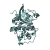
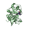
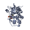
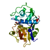
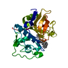
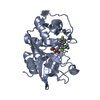
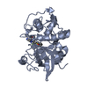
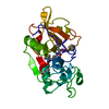
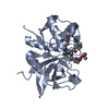
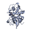
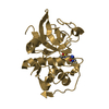
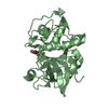
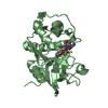
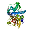
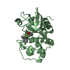
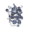
| Entry | Database: PDB / ID: 2r9o | ||||||
|---|---|---|---|---|---|---|---|
| Title | Cathepsin S complexed with Compound 8 | ||||||
 Components Components | Cathepsin S | ||||||
 Keywords Keywords | HYDROLASE / CATHEPSIN / PROTEASE / Glycoprotein / Lysosome / Polymorphism / Thiol protease / Zymogen | ||||||
| Function / homology |  Function and homology informationcathepsin S / basement membrane disassembly / positive regulation of cation channel activity / antigen processing and presentation of peptide antigen / endolysosome lumen / response to acidic pH / cellular response to thyroid hormone stimulus / Trafficking and processing of endosomal TLR / proteoglycan binding / Assembly of collagen fibrils and other multimeric structures ...cathepsin S / basement membrane disassembly / positive regulation of cation channel activity / antigen processing and presentation of peptide antigen / endolysosome lumen / response to acidic pH / cellular response to thyroid hormone stimulus / Trafficking and processing of endosomal TLR / proteoglycan binding / Assembly of collagen fibrils and other multimeric structures / toll-like receptor signaling pathway / cysteine-type endopeptidase activator activity involved in apoptotic process / fibronectin binding / antigen processing and presentation / collagen catabolic process / extracellular matrix disassembly / laminin binding / phagocytic vesicle / collagen binding / MHC class II antigen presentation / Degradation of the extracellular matrix / lysosomal lumen / proteolysis involved in protein catabolic process / positive regulation of apoptotic signaling pathway / Endosomal/Vacuolar pathway / protein processing / antigen processing and presentation of exogenous peptide antigen via MHC class II / late endosome / tertiary granule lumen / collagen-containing extracellular matrix / adaptive immune response / ficolin-1-rich granule lumen / lysosome / immune response / cysteine-type endopeptidase activity / serine-type endopeptidase activity / intracellular membrane-bounded organelle / Neutrophil degranulation / proteolysis / extracellular space / extracellular region Function and homology informationcathepsin S / basement membrane disassembly / positive regulation of cation channel activity / antigen processing and presentation of peptide antigen / endolysosome lumen / response to acidic pH / cellular response to thyroid hormone stimulus / Trafficking and processing of endosomal TLR / proteoglycan binding / Assembly of collagen fibrils and other multimeric structures ...cathepsin S / basement membrane disassembly / positive regulation of cation channel activity / antigen processing and presentation of peptide antigen / endolysosome lumen / response to acidic pH / cellular response to thyroid hormone stimulus / Trafficking and processing of endosomal TLR / proteoglycan binding / Assembly of collagen fibrils and other multimeric structures / toll-like receptor signaling pathway / cysteine-type endopeptidase activator activity involved in apoptotic process / fibronectin binding / antigen processing and presentation / collagen catabolic process / extracellular matrix disassembly / laminin binding / phagocytic vesicle / collagen binding / MHC class II antigen presentation / Degradation of the extracellular matrix / lysosomal lumen / proteolysis involved in protein catabolic process / positive regulation of apoptotic signaling pathway / Endosomal/Vacuolar pathway / protein processing / antigen processing and presentation of exogenous peptide antigen via MHC class II / late endosome / tertiary granule lumen / collagen-containing extracellular matrix / adaptive immune response / ficolin-1-rich granule lumen / lysosome / immune response / cysteine-type endopeptidase activity / serine-type endopeptidase activity / intracellular membrane-bounded organelle / Neutrophil degranulation / proteolysis / extracellular space / extracellular regionSimilarity search - Function | ||||||
| Biological species |  Homo sapiens (human) Homo sapiens (human) | ||||||
| Method | X-RAY DIFFRACTION / Resolution: 2 Å | ||||||
 Authors Authors | Ward, Y.D. / Emmanuel, M.J. / Thomson, D.S. / Liu, W. / Bekkali, Y. / Frye, L.L. / Girardot, M. / Morwick, T. / Young, E.R.R. / Zindell, R. ...Ward, Y.D. / Emmanuel, M.J. / Thomson, D.S. / Liu, W. / Bekkali, Y. / Frye, L.L. / Girardot, M. / Morwick, T. / Young, E.R.R. / Zindell, R. / Hrapchak, M. / DeTuri, M. / White, A. / Crane, K.M. / White, D.M. / Wang, Y. / Hao, M.-H. / Grygon, C.A. / Labadia, M.E. / Wildeson, J. / Freeman, D. / Nelson, R. / Capolino, A. / Peterson, J.D. / Raymond, E.L. / Brown, M.L. / Spero, D.M. | ||||||
 Citation Citation | Journal: to be published Title: Design and Synthesis of Reversible Inhibitors of Cathepsin S: alpha,alpha-Disubstitution at the P1 Residue Provides Potent Inhibitors in Cellular Assays and In Vivo Models of Antigen Presentation Authors: Ward, Y.D. / Emmanuel, M.J. / Thomson, D.S. / Liu, W. / Bekkali, Y. / Frye, L.L. / Girardot, M. / Morwick, T. / Young, E.R.R. / Zindell, R. / Hrapchak, M. / DeTuri, M. / White, A. / Crane, K. ...Authors: Ward, Y.D. / Emmanuel, M.J. / Thomson, D.S. / Liu, W. / Bekkali, Y. / Frye, L.L. / Girardot, M. / Morwick, T. / Young, E.R.R. / Zindell, R. / Hrapchak, M. / DeTuri, M. / White, A. / Crane, K.M. / White, D.M. / Wang, Y. / Hao, M.-H. / Grygon, C.A. / Labadia, M.E. / Wildeson, J. / Freeman, D. / Nelson, R. / Capolino, A. / Peterson, J.D. / Raymond, E.L. / Brown, M.L. / Spero, D.M. | ||||||
| History |
|
- Structure visualization
| Structure viewer | Molecule: MolmilJmol/JSmol |
|---|
- Downloads & links
-Download
| PDBx/mmCIF format | 2r9o.cif.gz | 102.9 KB | Display | PDBx/mmCIF format |
|---|---|---|---|---|
| PDB format | pdb2r9o.ent.gz | 79.5 KB | Display | PDB format |
| PDBx/mmJSON format | 2r9o.json.gz | Tree view | PDBx/mmJSON format | |
| Others |  Other downloads Other downloads |
-Validation report
| Arichive directory | https://data.pdbj.org/pub/pdb/validation_reports/r9/2r9oftp://data.pdbj.org/pub/pdb/validation_reports/r9/2r9o | HTTPS FTP |
|---|
-Related structure data
| Related structure data |  2r9mC  2r9nC  1ms6S C: citing same article ( S: Starting model for refinement |
|---|---|
| Similar structure data |


-Links
 PDBj
PDBj


- Assembly
| Deposited unit | 
| ||||||||
|---|---|---|---|---|---|---|---|---|---|
| 1 | 
| ||||||||
| 2 | 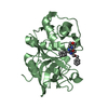
| ||||||||
| Unit cell |
| ||||||||
| Components on special symmetry positions |
|
-Components
| #1: Protein | Mass: 24697.670 Da / Num. of mol.: 2 Source method: isolated from a genetically manipulated source Source: (gene. exp.) Homo sapiens (human) / Gene: CTSS / Production host:  Escherichia coli (E. coli) / References: UniProt: P25774, cathepsin S Escherichia coli (E. coli) / References: UniProt: P25774, cathepsin S#2: Chemical |   Mass: 456.578 Da / Num. of mol.: 2 / Source method: obtained synthetically / Formula: C25H36N4O4 Mass: 456.578 Da / Num. of mol.: 2 / Source method: obtained synthetically / Formula: C25H36N4O4#3: Water | ChemComp-HOH / | Water Mass: 18.015 Da / Num. of mol.: 337 / Source method: isolated from a natural source / Formula: H2O Mass: 18.015 Da / Num. of mol.: 337 / Source method: isolated from a natural source / Formula: H2O |
|---|
-Experimental details
-Experiment
| Experiment | Method: X-RAY DIFFRACTION / Number of used crystals: 1 |
|---|
- Sample preparation
| Crystal | Density Matthews: 2.77 Å3/Da / Density % sol: 55.6 % |
|---|---|
| Crystal grow | Temperature: 298 K / Method: vapor diffusion, hanging drop / pH: 4.6 Details: 100mM NaOAc, 18-35% PEG MME (500-8000), 2 M (NH4)2SO4 with or without 15% glycerol, pH 4.6, vapor diffusion, hanging drop, temperature 298K |
-Data collection
| Diffraction | Mean temperature: 100 K |
|---|---|
| Diffraction source | Source: ROTATING ANODE / Type: RIGAKU RU200 / Wavelength: 1.5418 Å |
| Detector | Type: RIGAKU RAXIS IIC / Detector: IMAGE PLATE / Details: Osmic focussing mirrors |
| Radiation | Protocol: SINGLE WAVELENGTH / Monochromatic (M) / Laue (L): M / Scattering type: x-ray |
| Radiation wavelength | Wavelength: 1.5418 Å / Relative weight: 1 |
| Reflection | Resolution: 2→50 Å / Num. obs: 38017 / % possible obs: 99 % / Observed criterion σ(F): 0 / Observed criterion σ(I): 0 / Redundancy: 7.5 % / Rsym value: 0.075 |
- Processing
| Software |
| ||||||||||||||||||||||||||||
|---|---|---|---|---|---|---|---|---|---|---|---|---|---|---|---|---|---|---|---|---|---|---|---|---|---|---|---|---|---|
| Refinement | Starting model: pdb entry 1MS6 Resolution: 2→50 Å / Cross valid method: THROUGHOUT / σ(F): 0 / σ(I): 0 / Stereochemistry target values: Engh & Huber
| ||||||||||||||||||||||||||||
| Displacement parameters | Biso mean: 15.229 Å2 | ||||||||||||||||||||||||||||
| Refinement step | Cycle: LAST / Resolution: 2→50 Å
| ||||||||||||||||||||||||||||
| Refine LS restraints |
|
