 Movie
Movie Controller

[English] 日本語
 Yorodumi
- PDB-1mj8: High Resolution Crystal Structure Of The Fab Fragment of The Este... -
+ Open data

- Basic information
| Entry | Database: PDB / ID: 1mj8 | ||||||
|---|---|---|---|---|---|---|---|
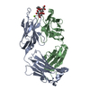
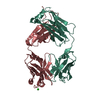
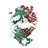
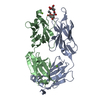
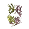
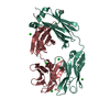
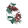
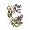
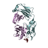
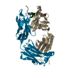
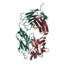
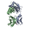
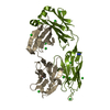
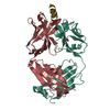
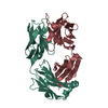
| Title | High Resolution Crystal Structure Of The Fab Fragment of The Esterolytic Antibody MS6-126 | ||||||
 Components Components | (IMMUNOGLOBULIN MS6-126) x 2 | ||||||
 Keywords Keywords |  IMMUNE SYSTEM / CATALYTIC ANTIBODY / ESTER HYDROLYSIS / ESTEROLYTIC / FAB / IMMUNOGLOBULIN IMMUNE SYSTEM / CATALYTIC ANTIBODY / ESTER HYDROLYSIS / ESTEROLYTIC / FAB / IMMUNOGLOBULIN | ||||||
| Function / homology | Immunoglobulins / Immunoglobulin-like / Sandwich / Mainly Beta / PHOSPHATE ION Function and homology information Function and homology information | ||||||
| Biological species |  Mus musculus (house mouse) Mus musculus (house mouse) | ||||||
| Method | X-RAY DIFFRACTION / SYNCHROTRON / MOLECULAR REPLACEMENT / Resolution: 1.75 Å | ||||||
 Authors Authors | Ruzheinikov, S.N. / Muranova, T.A. / Sedelnikova, S.E. / Partridge, L.J. / Blackburn, G.M. / Murray, I.A. / Kakinuma, H. / Takashi, N. / Shimazaki, K. / Sun, J. ...Ruzheinikov, S.N. / Muranova, T.A. / Sedelnikova, S.E. / Partridge, L.J. / Blackburn, G.M. / Murray, I.A. / Kakinuma, H. / Takashi, N. / Shimazaki, K. / Sun, J. / Nishi, Y. / Rice, D.W. | ||||||
 Citation Citation | Journal: J.Mol.Biol. / Year: 2003 Title: High-resolution crystal structure of the Fab-fragments of a family of mouse catalytic antibodies with esterase activity Authors: Ruzheinikov, S.N. / Muranova, T.A. / Sedelnikova, S.E. / Partridge, L.J. / Blackburn, G.M. / Murray, I.A. / Kakinuma, H. / Takashi, N. / Shimazaki, K. / Sun, J. / Nishi, Y. / Rice, D.W. #1: Journal: Acta Crystallogr.,Sect.D / Year: 2001Title: The preparation and crystallization of Fab fragments of a family of mouse esterolytic catalytic antibodies and their complexes with a transition-state analogue Authors: Muranova, T.A. / Ruzheinikov, S.N. / Sedelnikova, S.E. / Moir, A. / Partridge, L.J. / Kakinuma, H. / Takashi, N. / Shimazaki, K. / Sun, J. / Nishi, Y. / Rice, D.W. | ||||||
| History |
| ||||||
| Remark 999 | sequence an appropriate sequence database reference was not available at the time of processing. |
- Structure visualization
| Structure viewer | Molecule: MolmilJmol/JSmol |
|---|
- Downloads & links
-Download
| PDBx/mmCIF format | 1mj8.cif.gz | 111.8 KB | Display | PDBx/mmCIF format |
|---|---|---|---|---|
| PDB format | pdb1mj8.ent.gz | 88.4 KB | Display | PDB format |
| PDBx/mmJSON format | 1mj8.json.gz | Tree view | PDBx/mmJSON format | |
| Others |  Other downloads Other downloads |
-Validation report
| Arichive directory | https://data.pdbj.org/pub/pdb/validation_reports/mj/1mj8ftp://data.pdbj.org/pub/pdb/validation_reports/mj/1mj8 | HTTPS FTP |
|---|
-Related structure data
| Related structure data | 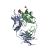 1mh5SC  1mieC  1mj7C  1mjjC 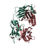 1mjuC S: Starting model for refinement C: citing same article ( |
|---|---|
| Similar structure data |


-Links
 PDBj
PDBj

- Assembly
| Deposited unit | 
| ||||||||
|---|---|---|---|---|---|---|---|---|---|
| 1 |
| ||||||||
| Unit cell |
|
-Components
| #1: Antibody | Mass: 24205.926 Da / Num. of mol.: 1 / Fragment: Fab Fragment, LIGHT CHAIN Source method: isolated from a genetically manipulated source Source: (gene. exp.) Mus musculus (house mouse) / Description: fusing of mouse splenocytes with myeloma cell / Cell line: hybridoma / Production host: Mus musculus (house mouse) | ||||
|---|---|---|---|---|---|
| #2: Antibody | Mass: 24685.738 Da / Num. of mol.: 1 / Fragment: Fab Fragment, HEAVY CHAIN Source method: isolated from a genetically manipulated source Source: (gene. exp.) Mus musculus (house mouse) / Description: fusing of mouse splenocytes with myeloma cell / Cell line: hybridoma / Production host: Mus musculus (house mouse) | ||||
| #3: Chemical | ChemComp-PO4 / Phosphate  Mass: 94.971 Da / Num. of mol.: 5 / Source method: obtained synthetically / Formula: PO4 Mass: 94.971 Da / Num. of mol.: 5 / Source method: obtained synthetically / Formula: PO4#4: Chemical | Glycerol  Mass: 92.094 Da / Num. of mol.: 2 / Source method: obtained synthetically / Formula: C3H8O3 Mass: 92.094 Da / Num. of mol.: 2 / Source method: obtained synthetically / Formula: C3H8O3#5: Water | ChemComp-HOH / | Water Mass: 18.015 Da / Num. of mol.: 653 / Source method: isolated from a natural source / Formula: H2O Mass: 18.015 Da / Num. of mol.: 653 / Source method: isolated from a natural source / Formula: H2O |
-Experimental details
-Experiment
| Experiment | Method: X-RAY DIFFRACTION / Number of used crystals: 1 |
|---|
- Sample preparation
| Crystal | Density Matthews: 2.27 Å3/Da / Density % sol: 45.85 % | ||||||||||||||||||||||||||||||||||||||||||
|---|---|---|---|---|---|---|---|---|---|---|---|---|---|---|---|---|---|---|---|---|---|---|---|---|---|---|---|---|---|---|---|---|---|---|---|---|---|---|---|---|---|---|---|
| Crystal grow | Temperature: 290 K / Method: vapor diffusion, hanging drop / pH: 8.5 Details: Ammonium phosphate, Tris-HCl, pH 8.5, VAPOR DIFFUSION, HANGING DROP, temperature 290K | ||||||||||||||||||||||||||||||||||||||||||
| Crystal grow | *PLUS Method: vapor diffusion, hanging drop | ||||||||||||||||||||||||||||||||||||||||||
| Components of the solutions | *PLUS
|
-Data collection
| Diffraction | Mean temperature: 100 K |
|---|---|
| Diffraction source | Source: SYNCHROTRON / Site: SRS  / Beamline: PX9.6 / Wavelength: 0.87 Å / Beamline: PX9.6 / Wavelength: 0.87 Å |
| Detector | Type: ADSC QUANTUM 4 / Detector: CCD / Date: Sep 6, 2000 / Details: mirrors |
| Radiation | Monochromator: Si 111 / Protocol: SINGLE WAVELENGTH / Monochromatic (M) / Laue (L): M / Scattering type: x-ray |
| Radiation wavelength | Wavelength: 0.87 Å / Relative weight: 1 |
| Reflection | Resolution: 1.75→10 Å / Num. all: 39166 / Num. obs: 39166 / % possible obs: 89.1 % / Observed criterion σ(F): 0 / Observed criterion σ(I): -3 / Redundancy: 3.5 % / Rmerge(I) obs: 0.037 / Net I/σ(I): 31 |
| Reflection shell | Resolution: 1.75→1.78 Å / Rmerge(I) obs: 0.183 / Mean I/σ(I) obs: 4.7 / % possible all: 58.4 |
| Reflection | *PLUS |
| Reflection shell | *PLUS % possible obs: 58.4 % |
- Processing
| Software |
| ||||||||||||||||||||
|---|---|---|---|---|---|---|---|---|---|---|---|---|---|---|---|---|---|---|---|---|---|
| Refinement | Method to determine structure: MOLECULAR REPLACEMENT Starting model: PDB ENTRY 1mh5 Resolution: 1.75→10 Å / Isotropic thermal model: Isotropic / Cross valid method: THROUGHOUT / σ(F): 0 / Stereochemistry target values: Engh & Huber
| ||||||||||||||||||||
| Displacement parameters | Biso mean: 24.8 Å2 | ||||||||||||||||||||
| Refinement step | Cycle: LAST / Resolution: 1.75→10 Å
| ||||||||||||||||||||
| Refine LS restraints |
| ||||||||||||||||||||
| Xplor file |
| ||||||||||||||||||||
| Refinement | *PLUS % reflection Rfree: 5 % | ||||||||||||||||||||
| Solvent computation | *PLUS | ||||||||||||||||||||
| Displacement parameters | *PLUS |
