National Institutes of Health/National Institute of General Medical Sciences (NIH/NIGMS)
R01GM120553
United States
Citation
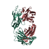
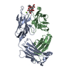
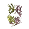
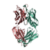
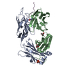
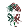
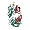
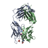
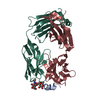
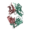
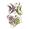
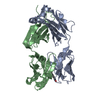
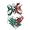
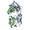
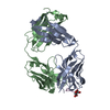
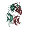
Journal: Nat Struct Mol Biol / Year: 2019 Title: An antibody against the F glycoprotein inhibits Nipah and Hendra virus infections. Authors: Ha V Dang / Yee-Peng Chan / Young-Jun Park / Joost Snijder / Sofia Cheliout Da Silva / Bang Vu / Lianying Yan / Yan-Ru Feng / Barry Rockx / Thomas W Geisbert / Chad E Mire / Christopher C ...Authors: Ha V Dang / Yee-Peng Chan / Young-Jun Park / Joost Snijder / Sofia Cheliout Da Silva / Bang Vu / Lianying Yan / Yan-Ru Feng / Barry Rockx / Thomas W Geisbert / Chad E Mire / Christopher C Broder / David Veesler / Abstract: Nipah virus (NiV) and Hendra virus (HeV) are zoonotic henipaviruses (HNVs) responsible for outbreaks of encephalitis and respiratory illness with fatality rates of 50-100%. No vaccines or licensed ...Nipah virus (NiV) and Hendra virus (HeV) are zoonotic henipaviruses (HNVs) responsible for outbreaks of encephalitis and respiratory illness with fatality rates of 50-100%. No vaccines or licensed therapeutics currently exist to protect humans against NiV or HeV. HNVs enter host cells by fusing the viral and cellular membranes via the concerted action of the attachment (G) and fusion (F) glycoproteins, the main targets of the humoral immune response. Here, we describe the isolation and humanization of a potent monoclonal antibody cross-neutralizing NiV and HeV. Cryo-electron microscopy, triggering and fusion studies show the antibody binds to a prefusion-specific quaternary epitope, conserved in NiV F and HeV F glycoproteins, and prevents membrane fusion and viral entry. This work supports the importance of the HNV prefusion F conformation for eliciting a robust immune response and paves the way for using this antibody for prophylaxis and post-exposure therapy with NiV- and HeV-infected individuals.
Resolution: 1.483→68.6 Å / Cor.coef. Fo:Fc: 0.979 / Cor.coef. Fo:Fc free: 0.97 / SU B: 3.593 / SU ML: 0.055 / SU R Cruickshank DPI: 0.0647 / Cross valid method: THROUGHOUT / σ(F): 0 / ESU R: 0.065 / ESU R Free: 0.06 Details: HYDROGENS HAVE BEEN ADDED IN THE RIDING POSITIONS U VALUES : WITH TLS ADDED
Rfactor
Num. reflection
% reflection
Selection details
Rfree
0.1733
4238
5 %
RANDOM
Rwork
0.1372
-
-
-
obs
0.139
79965
99.61 %
-
Solvent computation
Ion probe radii: 0.8 Å / Shrinkage radii: 0.8 Å / VDW probe radii: 1.2 Å
In the structure databanks used in Yorodumi, some data are registered as the other names, "COVID-19 virus" and "2019-nCoV". Here are the details of the virus and the list of structure data.
Jan 31, 2019. EMDB accession codes are about to change! (news from PDBe EMDB page)
EMDB accession codes are about to change! (news from PDBe EMDB page)
The allocation of 4 digits for EMDB accession codes will soon come to an end. Whilst these codes will remain in use, new EMDB accession codes will include an additional digit and will expand incrementally as the available range of codes is exhausted. The current 4-digit format prefixed with “EMD-” (i.e. EMD-XXXX) will advance to a 5-digit format (i.e. EMD-XXXXX), and so on. It is currently estimated that the 4-digit codes will be depleted around Spring 2019, at which point the 5-digit format will come into force.
The EM Navigator/Yorodumi systems omit the EMD- prefix.
Related info.:Q: What is EMD? / ID/Accession-code notation in Yorodumi/EM Navigator
Yorodumi is a browser for structure data from EMDB, PDB, SASBDB, etc.
This page is also the successor to EM Navigator detail page, and also detail information page/front-end page for Omokage search.
The word "yorodu" (or yorozu) is an old Japanese word meaning "ten thousand". "mi" (miru) is to see.
Related info.:EMDB / PDB / SASBDB / Comparison of 3 databanks / Yorodumi Search / Aug 31, 2016. New EM Navigator & Yorodumi / Yorodumi Papers / Jmol/JSmol / Function and homology information / Changes in new EM Navigator and Yorodumi
 Movie
Movie Controller

 Yorodumi
 Open data
 Basic information
 Components
 Keywords
 IMMUNE SYSTEM /
  Function and homology information
 Authors
 United States, 1items
  Citation
 Structure visualization
 Downloads & links
 Other downloads
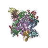


 PDBj
PDBj

 Assembly
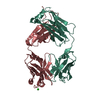

 Mass: 35.453 Da / Num. of mol.: 1 / Source method: obtained synthetically / Formula: Cl
 Mass: 18.015 Da / Num. of mol.: 557 / Source method: isolated from a natural source / Formula: H2O
 Sample preparation
 Processing