 Movie
Movie Controller

[English] 日本語
 Yorodumi
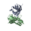
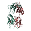
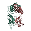
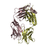
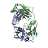
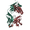
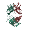
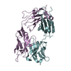
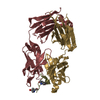
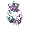
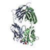
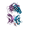
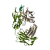
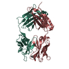
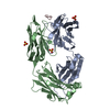
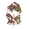
- PDB-1fvd: X-RAY STRUCTURES OF THE ANTIGEN-BINDING DOMAINS FROM THREE VARIAN... -
+ Open data

- Basic information
| Entry | Database: PDB / ID: 1fvd | ||||||
|---|---|---|---|---|---|---|---|
| Title | X-RAY STRUCTURES OF THE ANTIGEN-BINDING DOMAINS FROM THREE VARIANTS OF HUMANIZED ANTI-P185-HER2 ANTIBODY 4D5 AND COMPARISON WITH MOLECULAR MODELING | ||||||
 Components Components |
| ||||||
 Keywords Keywords |  IMMUNOGLOBULIN IMMUNOGLOBULIN | ||||||
| Function / homology | Immunoglobulins / Immunoglobulin-like / Sandwich / Mainly Beta / : / :  Function and homology information Function and homology information | ||||||
| Biological species |  Homo sapiens (human) Homo sapiens (human) | ||||||
| Method | X-RAY DIFFRACTION / Resolution: 2.5 Å | ||||||
 Authors Authors | Eigenbrot, C. / Presta, L. / Randal, M. / Kossiakoff, A.A. | ||||||
 Citation Citation | Journal: J.Mol.Biol. / Year: 1993 Title: X-ray structures of the antigen-binding domains from three variants of humanized anti-p185HER2 antibody 4D5 and comparison with molecular modeling. Authors: Eigenbrot, C. / Randal, M. / Presta, L. / Carter, P. / Kossiakoff, A.A. #1: Journal: Proc.Natl.Acad.Sci.USA / Year: 1992Title: Humanization of an Anti-P185-Her2 Antibody for Human Cancer Therapy Authors: Carter, P. / Presta, L. / Gorman, C.M. / Ridgway, J.B. / Henner, D. / Wong, W.L.T. / Rowland, A.M. / Kotts, C. / Carver, M.E. / Shepard, H.M. | ||||||
| History |
|
- Structure visualization
| Structure viewer | Molecule: MolmilJmol/JSmol |
|---|
- Downloads & links
-Download
| PDBx/mmCIF format | 1fvd.cif.gz | 174.2 KB | Display | PDBx/mmCIF format |
|---|---|---|---|---|
| PDB format | pdb1fvd.ent.gz | 145.5 KB | Display | PDB format |
| PDBx/mmJSON format | 1fvd.json.gz | Tree view | PDBx/mmJSON format | |
| Others |  Other downloads Other downloads |
-Validation report
| Arichive directory | https://data.pdbj.org/pub/pdb/validation_reports/fv/1fvdftp://data.pdbj.org/pub/pdb/validation_reports/fv/1fvd | HTTPS FTP |
|---|
-Related structure data


-Links
 PDBj
PDBj

- Assembly
| Deposited unit | 
| ||||||||
|---|---|---|---|---|---|---|---|---|---|
| 1 | 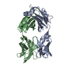
| ||||||||
| 2 | 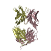
| ||||||||
| Unit cell |
| ||||||||
| Atom site foot note | 1: RESIDUES 8, 95, 141 OF CHAINS A AND C AND RESIDUES 154 AND 156 OF CHAINS B AND D ARE CIS PROLINES. 2: 127 ATOMS OF THE FINAL MODEL WERE ASSIGNED AN OCCUPANCY OF ZERO. THEY ARE IN CDR-H3, THE C-TERMINAL REGIONS, AND A CONSTANT DOMAIN LOOP REGION. | ||||||||
| Noncrystallographic symmetry (NCS) | NCS oper: (Code: given Matrix: (-0.99983, 0.0176, -0.00577), Vector : Details | THE TRANSFORMATION PRESENTED ON *MTRIX* RECORDS BELOW WILL YIELD APPROXIMATE COORDINATES FOR CHAINS *A* AND *B* WHEN APPLIED TO CHAINS *C* AND *D*. | |
-Components
| #1: Antibody | Mass: 23431.973 Da / Num. of mol.: 2 Source method: isolated from a genetically manipulated source Source: (gene. exp.) Homo sapiens (human) / Production host:  Escherichia coli (E. coli) / References: EMBL: X95750 Escherichia coli (E. coli) / References: EMBL: X95750#2: Antibody | Mass: 23722.617 Da / Num. of mol.: 2 Source method: isolated from a genetically manipulated source Source: (gene. exp.) Homo sapiens (human) / Production host: Escherichia coli (E. coli) / References: EMBL: Y14735#3: Water | ChemComp-HOH / | Water Mass: 18.015 Da / Num. of mol.: 199 / Source method: isolated from a natural source / Formula: H2O Mass: 18.015 Da / Num. of mol.: 199 / Source method: isolated from a natural source / Formula: H2OSequence details | THE RESIDUE NUMBERING IS SEQUENTIAL WITHIN EACH CHAIN. THE SEQUENTIAL NUMBERING OF THE LIGHT CHAIN ...THE RESIDUE NUMBERING IS SEQUENTIAL | |
|---|
-Experimental details
-Experiment
| Experiment | Method: X-RAY DIFFRACTION |
|---|
- Sample preparation
| Crystal | Density Matthews: 2.55 Å3/Da / Density % sol: 51.7 % | ||||||||||||||||||||
|---|---|---|---|---|---|---|---|---|---|---|---|---|---|---|---|---|---|---|---|---|---|
| Crystal grow | *PLUS Method: vapor diffusion, hanging drop | ||||||||||||||||||||
| Components of the solutions | *PLUS
|
-Data collection
| Radiation | Scattering type: x-ray |
|---|---|
| Radiation wavelength | Relative weight: 1 |
| Reflection | *PLUS Highest resolution: 2.5 Å / Lowest resolution: 15 Å / Num. obs: 27905 / Observed criterion σ(I): 0 / Num. measured all: 50304 / Rmerge(I) obs: 0.119 |
- Processing
| Software |
| ||||||||||||||||||||||||||||||||||||||||||||||||||||||||||||
|---|---|---|---|---|---|---|---|---|---|---|---|---|---|---|---|---|---|---|---|---|---|---|---|---|---|---|---|---|---|---|---|---|---|---|---|---|---|---|---|---|---|---|---|---|---|---|---|---|---|---|---|---|---|---|---|---|---|---|---|---|---|
| Refinement | Resolution: 2.5→10 Å / Rfactor Rwork: 0.179 / Rfactor obs: 0.179 / σ(F): 2 | ||||||||||||||||||||||||||||||||||||||||||||||||||||||||||||
| Refinement step | Cycle: LAST / Resolution: 2.5→10 Å
| ||||||||||||||||||||||||||||||||||||||||||||||||||||||||||||
| Refine LS restraints |
| ||||||||||||||||||||||||||||||||||||||||||||||||||||||||||||
| Refinement | *PLUS Highest resolution: 2.5 Å / Lowest resolution: 10 Å / Num. reflection obs: 26210 / σ(F): 2 / Rfactor obs: 0.179 | ||||||||||||||||||||||||||||||||||||||||||||||||||||||||||||
| Solvent computation | *PLUS | ||||||||||||||||||||||||||||||||||||||||||||||||||||||||||||
| Displacement parameters | *PLUS Biso mean: 18.9 Å2 | ||||||||||||||||||||||||||||||||||||||||||||||||||||||||||||
| Refine LS restraints | *PLUS
|
