 Movie
Movie Controller

[English] 日本語
 Yorodumi
- PDB-6bzu: Structure of the Hepatitis C virus envelope glycoprotein E2 antig... -
+ Open data

- Basic information
| Entry | Database: PDB / ID: 6bzu | |||||||||
|---|---|---|---|---|---|---|---|---|---|---|
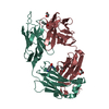
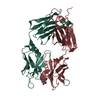
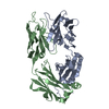
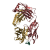
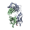
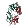
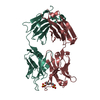
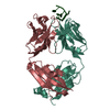
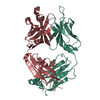
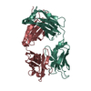
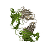
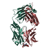
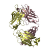
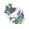
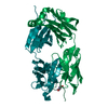
| Title | Structure of the Hepatitis C virus envelope glycoprotein E2 antigenic region 412-423 bound to the broadly neutralizing antibody 19B3 | |||||||||
 Components Components |
| |||||||||
 Keywords Keywords |  IMMUNE SYSTEM / Hepatitis C virus / antibodies / broadly neutralizing antibodies / rational vaccine design / AS412 / E2 / VIRAL PROTEIN IMMUNE SYSTEM / Hepatitis C virus / antibodies / broadly neutralizing antibodies / rational vaccine design / AS412 / E2 / VIRAL PROTEIN | |||||||||
| Function / homology |  Function and homology information Function and homology informationpositive regulation of hexokinase activity / modulation by virus of host cellular process / translocation of peptides or proteins into host cell cytoplasm / Toll-like receptor 2 binding / viral capsid assembly / adhesion receptor-mediated virion attachment to host cell / TBC/RABGAPs / hepacivirin / host cell mitochondrial membrane / host cell lipid droplet ...positive regulation of hexokinase activity / modulation by virus of host cellular process / translocation of peptides or proteins into host cell cytoplasm / Toll-like receptor 2 binding / viral capsid assembly / adhesion receptor-mediated virion attachment to host cell / TBC/RABGAPs / hepacivirin / host cell mitochondrial membrane / host cell lipid droplet / symbiont-mediated suppression of host TRAF-mediated signal transduction / transformation of host cell by virus / positive regulation of cytokinesis / symbiont-mediated perturbation of host cell cycle G1/S transition checkpoint / negative regulation of protein secretion / symbiont-mediated suppression of host JAK-STAT cascade via inhibition of STAT1 activity / symbiont-mediated suppression of host cytoplasmic pattern recognition receptor signaling pathway via inhibition of MAVS activity / endoplasmic reticulum-Golgi intermediate compartment membrane / SH3 domain binding / kinase binding / : / nucleoside-triphosphate phosphatase / protein complex oligomerization / monoatomic ion channel activity / clathrin-dependent endocytosis of virus by host cell / viral nucleocapsid / Hydrolases; Acting on peptide bonds (peptidases); Cysteine endopeptidases / entry receptor-mediated virion attachment to host cell / RNA helicase activity / host cell endoplasmic reticulum membrane / host cell perinuclear region of cytoplasm / RNA helicase / induction by virus of host autophagy / ribonucleoprotein complex / RNA-directed RNA polymerase / viral RNA genome replication / cysteine-type endopeptidase activity / RNA-dependent RNA polymerase activity / serine-type endopeptidase activity / fusion of virus membrane with host endosome membrane / viral envelope / symbiont-mediated suppression of host type I interferon-mediated signaling pathway / host cell nucleus / host cell plasma membrane / virion membrane / structural molecule activity / negative regulation of transcription by RNA polymerase II / ATP hydrolysis activity / proteolysis / RNA binding / zinc ion binding / ATP bindingSimilarity search - Function | |||||||||
| Biological species |  Mus musculus (house mouse) Mus musculus (house mouse) Hepatitis C virus Hepatitis C virus | |||||||||
| Method | X-RAY DIFFRACTION / SYNCHROTRON / MOLECULAR REPLACEMENT / Resolution: 2.7 Å | |||||||||
 Authors Authors | Tzarum, N. / Aleman, F. / Kong, L. / Wilson, I.A. / Law, M. | |||||||||
| Funding support |  United States, 2items United States, 2items
| |||||||||
 Citation Citation | Journal: Proc. Natl. Acad. Sci. U.S.A. / Year: 2018 Title: Immunogenetic and structural analysis of a class of HCV broadly neutralizing antibodies and their precursors. Authors: Aleman, F. / Tzarum, N. / Kong, L. / Nagy, K. / Zhu, J. / Wilson, I.A. / Law, M. | |||||||||
| History |
|
- Structure visualization
| Structure viewer | Molecule: MolmilJmol/JSmol |
|---|
- Downloads & links
-Download
| PDBx/mmCIF format | 6bzu.cif.gz | 340.2 KB | Display | PDBx/mmCIF format |
|---|---|---|---|---|
| PDB format | pdb6bzu.ent.gz | 275.4 KB | Display | PDB format |
| PDBx/mmJSON format | 6bzu.json.gz | Tree view | PDBx/mmJSON format | |
| Others |  Other downloads Other downloads |
-Validation report
| Arichive directory | https://data.pdbj.org/pub/pdb/validation_reports/bz/6bzuftp://data.pdbj.org/pub/pdb/validation_reports/bz/6bzu | HTTPS FTP |
|---|
-Related structure data
| Related structure data | 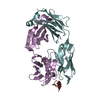 6bzvC  6bzwC  6bzyC  4g6aS S: Starting model for refinement C: citing same article ( |
|---|---|
| Similar structure data |


-Links
 PDBj
PDBj


- Assembly
| Deposited unit | 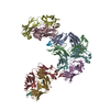
| ||||||||
|---|---|---|---|---|---|---|---|---|---|
| 1 | 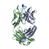
| ||||||||
| 2 | 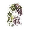
| ||||||||
| 3 | 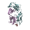
| ||||||||
| 4 | 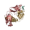
| ||||||||
| Unit cell |
|
-Components
| #1: Antibody | Mass: 23894.594 Da / Num. of mol.: 4 Source method: isolated from a genetically manipulated source Source: (gene. exp.) Mus musculus (house mouse) / Production host:  Homo sapiens (human) Homo sapiens (human)#2: Antibody | Mass: 23781.248 Da / Num. of mol.: 4 Source method: isolated from a genetically manipulated source Source: (gene. exp.) Mus musculus (house mouse) / Production host: Homo sapiens (human)#3: Protein/peptide | Mass: 1554.710 Da / Num. of mol.: 4 / Source method: obtained synthetically / Source: (synth.) Hepatitis C virus / References: UniProt: P27958*PLUS#4: Water | ChemComp-HOH / | Water Mass: 18.015 Da / Num. of mol.: 220 / Source method: isolated from a natural source / Formula: H2O Mass: 18.015 Da / Num. of mol.: 220 / Source method: isolated from a natural source / Formula: H2O |
|---|
-Experimental details
-Experiment
| Experiment | Method: X-RAY DIFFRACTION / Number of used crystals: 1 |
|---|
- Sample preparation
| Crystal | Density Matthews: 2.79 Å3/Da / Density % sol: 55.96 % |
|---|---|
| Crystal grow | Temperature: 293 K / Method: vapor diffusion, sitting drop Details: 20% (w/v) PEG 3500, 0.2 M sodium-thicoyanate, pH 6.9 |
-Data collection
| Diffraction | Mean temperature: 100 K |
|---|---|
| Diffraction source | Source: SYNCHROTRON / Site: APS / Beamline: 23-ID-B / Wavelength: 1.0332 Å |
| Detector | Type: DECTRIS PILATUS3 S 6M / Detector: PIXEL / Date: Oct 15, 2015 |
| Radiation | Protocol: SINGLE WAVELENGTH / Monochromatic (M) / Laue (L): M / Scattering type: x-ray |
| Radiation wavelength | Wavelength: 1.0332 Å / Relative weight: 1 |
| Reflection | Resolution: 2.7→29.82 Å / Num. obs: 59853 / % possible obs: 98.6 % / Redundancy: 4.1 % / Net I/σ(I): 8.5 |
| Reflection shell | Resolution: 2.7→2.77 Å |
- Processing
| Software |
| |||||||||||||||||||||||||||||||||||||||||||||||||||||||||||||||||||||||||||||||||||||||||||||||||||||||||||||||||||||||||||||||||||||||||||||||||||||||||||||||||
|---|---|---|---|---|---|---|---|---|---|---|---|---|---|---|---|---|---|---|---|---|---|---|---|---|---|---|---|---|---|---|---|---|---|---|---|---|---|---|---|---|---|---|---|---|---|---|---|---|---|---|---|---|---|---|---|---|---|---|---|---|---|---|---|---|---|---|---|---|---|---|---|---|---|---|---|---|---|---|---|---|---|---|---|---|---|---|---|---|---|---|---|---|---|---|---|---|---|---|---|---|---|---|---|---|---|---|---|---|---|---|---|---|---|---|---|---|---|---|---|---|---|---|---|---|---|---|---|---|---|---|---|---|---|---|---|---|---|---|---|---|---|---|---|---|---|---|---|---|---|---|---|---|---|---|---|---|---|---|---|---|---|---|
| Refinement | Method to determine structure: MOLECULAR REPLACEMENT Starting model: 4G6A Resolution: 2.7→29.819 Å / SU ML: 0.47 / Cross valid method: FREE R-VALUE / σ(F): 1.34 / Phase error: 33.26
| |||||||||||||||||||||||||||||||||||||||||||||||||||||||||||||||||||||||||||||||||||||||||||||||||||||||||||||||||||||||||||||||||||||||||||||||||||||||||||||||||
| Solvent computation | Shrinkage radii: 0.9 Å / VDW probe radii: 1.11 Å | |||||||||||||||||||||||||||||||||||||||||||||||||||||||||||||||||||||||||||||||||||||||||||||||||||||||||||||||||||||||||||||||||||||||||||||||||||||||||||||||||
| Refinement step | Cycle: LAST / Resolution: 2.7→29.819 Å
| |||||||||||||||||||||||||||||||||||||||||||||||||||||||||||||||||||||||||||||||||||||||||||||||||||||||||||||||||||||||||||||||||||||||||||||||||||||||||||||||||
| Refine LS restraints |
| |||||||||||||||||||||||||||||||||||||||||||||||||||||||||||||||||||||||||||||||||||||||||||||||||||||||||||||||||||||||||||||||||||||||||||||||||||||||||||||||||
| LS refinement shell |
|
