 Movie
Movie Controller


+ Open data

- Basic information
| Entry | Database: PDB / ID: 1amk | ||||||
|---|---|---|---|---|---|---|---|
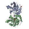
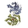
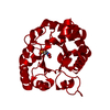
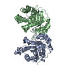
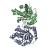
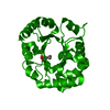
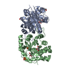
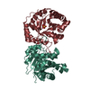
| Title | LEISHMANIA MEXICANA TRIOSE PHOSPHATE ISOMERASE | ||||||
 Components Components | TRIOSE PHOSPHATE ISOMERASE Triosephosphate isomerase Triosephosphate isomerase | ||||||
 Keywords Keywords | GLUCONEOGENESIS / TIM / 2-PG / PGA / FATTY ACID BIOSYNTHESIS | ||||||
| Function / homology |  Function and homology informationglycosome / glyceraldehyde-3-phosphate biosynthetic process / glycerol catabolic process / triose-phosphate isomerase / triose-phosphate isomerase activity / gluconeogenesis / glycolytic process / cytosol Function and homology informationglycosome / glyceraldehyde-3-phosphate biosynthetic process / glycerol catabolic process / triose-phosphate isomerase / triose-phosphate isomerase activity / gluconeogenesis / glycolytic process / cytosolSimilarity search - Function | ||||||
| Biological species |   Leishmania mexicana (eukaryote) Leishmania mexicana (eukaryote) | ||||||
| Method | X-RAY DIFFRACTION / MOLECULAR REPLACEMENT / Resolution: 1.83 Å | ||||||
 Authors Authors | Williams, J.C. / Wierenga, R. | ||||||
 Citation Citation | Journal: Protein Eng. / Year: 1999 Title: Structural and mutagenesis studies of leishmania triosephosphate isomerase: a point mutation can convert a mesophilic enzyme into a superstable enzyme without losing catalytic power. Authors: Williams, J.C. / Zeelen, J.P. / Neubauer, G. / Vriend, G. / Backmann, J. / Michels, P.A. / Lambeir, A.M. / Wierenga, R.K. #1: Journal: Eur.J.Biochem. / Year: 1994Title: Triose-Phosphate Isomerase of Leishmania Mexicana Mexicana. Cloning and Characterization of the Gene, Overexpression in Escherichia Coli and Analysis of the Protein Authors: Kohl, L. / Callens, M. / Wierenga, R.K. / Opperdoes, F.R. / Michels, P.A. | ||||||
| History |
|
- Structure visualization
| Structure viewer | Molecule: MolmilJmol/JSmol |
|---|
- Downloads & links
-Download
| PDBx/mmCIF format | 1amk.cif.gz | 63 KB | Display | PDBx/mmCIF format |
|---|---|---|---|---|
| PDB format | pdb1amk.ent.gz | 45.6 KB | Display | PDB format |
| PDBx/mmJSON format | 1amk.json.gz | Tree view | PDBx/mmJSON format | |
| Others |  Other downloads Other downloads |
-Validation report
| Arichive directory | https://data.pdbj.org/pub/pdb/validation_reports/am/1amkftp://data.pdbj.org/pub/pdb/validation_reports/am/1amk | HTTPS FTP |
|---|
-Related structure data
| Related structure data |  5timS S: Starting model for refinement |
|---|---|
| Similar structure data |


-Links
 PDBj
PDBj
- Assembly
| Deposited unit | 
| ||||||||
|---|---|---|---|---|---|---|---|---|---|
| 1 | 
| ||||||||
| Unit cell |
| ||||||||
| Components on special symmetry positions |
|
-Components
| #1: Protein | Triosephosphate isomerase Mass: 27209.223 Da / Num. of mol.: 1 Source method: isolated from a genetically manipulated source Source: (gene. exp.) Leishmania mexicana (eukaryote) / References: UniProt: P48499, triose-phosphate isomerase |
|---|---|
| #2: Chemical | ChemComp-PGA /   Mass: 156.031 Da / Num. of mol.: 1 Mass: 156.031 Da / Num. of mol.: 1Source method: isolated from a genetically manipulated source Formula: C2H5O6P |
| #3: Water | ChemComp-HOH / Water Mass: 18.015 Da / Num. of mol.: 112 / Source method: isolated from a natural source / Formula: H2O Mass: 18.015 Da / Num. of mol.: 112 / Source method: isolated from a natural source / Formula: H2O |
-Experimental details
-Experiment
| Experiment | Method: X-RAY DIFFRACTION / Number of used crystals: 1 |
|---|
- Sample preparation
| Crystal | Density Matthews: 2.5 Å3/Da / Density % sol: 51 % | ||||||||||||||||||||||||||||||||||||||||||||||||||||||||||||||||||||||||||||||||||||
|---|---|---|---|---|---|---|---|---|---|---|---|---|---|---|---|---|---|---|---|---|---|---|---|---|---|---|---|---|---|---|---|---|---|---|---|---|---|---|---|---|---|---|---|---|---|---|---|---|---|---|---|---|---|---|---|---|---|---|---|---|---|---|---|---|---|---|---|---|---|---|---|---|---|---|---|---|---|---|---|---|---|---|---|---|---|
| Crystal grow | pH: 5.85 Details: 100 MM ACETIC ACID/NAOH 4.5 20% PEG 6000 1 MM DTT, EDTA, 1 MM AZIDE, 5% ETHYLENE GLYCOL, 10 MM 2-PHOSPHOGLYCOLIC ACID. CRYSTAL WAS MOVED INTO 100 MM CITRATE PH 5.85 20 % PEG 6000, 1 MM DTT, ...Details: 100 MM ACETIC ACID/NAOH 4.5 20% PEG 6000 1 MM DTT, EDTA, 1 MM AZIDE, 5% ETHYLENE GLYCOL, 10 MM 2-PHOSPHOGLYCOLIC ACID. CRYSTAL WAS MOVED INTO 100 MM CITRATE PH 5.85 20 % PEG 6000, 1 MM DTT, 1 MM EDTA, 1 MM AZIDE BEFORE DATA COLLECTION. | ||||||||||||||||||||||||||||||||||||||||||||||||||||||||||||||||||||||||||||||||||||
| Crystal grow | *PLUS Method: vapor diffusion, hanging drop / pH: 7.5 | ||||||||||||||||||||||||||||||||||||||||||||||||||||||||||||||||||||||||||||||||||||
| Components of the solutions | *PLUS
|
-Data collection
| Diffraction | Mean temperature: 295 K |
|---|---|
| Diffraction source | Wavelength: 1.5418 |
| Detector | Type: ENRAF-NONIUS FAST / Detector: DIFFRACTOMETER / Date: Feb 22, 1996 |
| Radiation | Monochromatic (M) / Laue (L): M / Scattering type: x-ray |
| Radiation wavelength | Wavelength: 1.5418 Å / Relative weight: 1 |
| Reflection | Resolution: 1.8→22 Å / Num. obs: 22669 / % possible obs: 89.2 % / Redundancy: 5.5 % / Rmerge(I) obs: 0.048 / Net I/σ(I): 20.8 |
| Reflection shell | Resolution: 1.8→1.83 Å / Rmerge(I) obs: 0.116 / Mean I/σ(I) obs: 6 / % possible all: 84.3 |
| Reflection | *PLUS Lowest resolution: 22 Å / Num. measured all: 123706 / Rmerge(I) obs: 0.049 |
| Reflection shell | *PLUS % possible obs: 84.3 % / Rmerge(I) obs: 0.116 |
- Processing
| Software |
| ||||||||||||||||||||||||||||||||||||||||||||||||||
|---|---|---|---|---|---|---|---|---|---|---|---|---|---|---|---|---|---|---|---|---|---|---|---|---|---|---|---|---|---|---|---|---|---|---|---|---|---|---|---|---|---|---|---|---|---|---|---|---|---|---|---|
| Refinement | Method to determine structure: MOLECULAR REPLACEMENT Starting model: PDB ENTRY 5TIM Resolution: 1.83→22 Å / Isotropic thermal model: TNT BCORREL V1.0 / σ(F): 0 / Stereochemistry target values: TNT PROTGEO
| ||||||||||||||||||||||||||||||||||||||||||||||||||
| Solvent computation | Solvent model: BABINET / Bsol: 262.9 Å2 / ksol: 0.802 e/Å3 | ||||||||||||||||||||||||||||||||||||||||||||||||||
| Refinement step | Cycle: LAST / Resolution: 1.83→22 Å
| ||||||||||||||||||||||||||||||||||||||||||||||||||
| Refine LS restraints |
| ||||||||||||||||||||||||||||||||||||||||||||||||||
| Refinement | *PLUS Rfactor all: 0.107 / Rfactor Rwork: 0.107 | ||||||||||||||||||||||||||||||||||||||||||||||||||
| Solvent computation | *PLUS | ||||||||||||||||||||||||||||||||||||||||||||||||||
| Displacement parameters | *PLUS | ||||||||||||||||||||||||||||||||||||||||||||||||||
| Refine LS restraints | *PLUS
|
