 Movie
Movie Controller

[English] 日本語
 Yorodumi
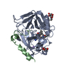
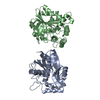
- PDB-1lyx: Plasmodium Falciparum Triosephosphate Isomerase (PfTIM)-Phosphogl... -
+ Open data

- Basic information
| Entry | Database: PDB / ID: 1lyx | ||||||
|---|---|---|---|---|---|---|---|
| Title | Plasmodium Falciparum Triosephosphate Isomerase (PfTIM)-Phosphoglycolate complex | ||||||
 Components Components | Triosephosphate Isomerase | ||||||
 Keywords Keywords | ISOMERASE / TIM barrels / beta-alpha barrels | ||||||
| Function / homology |  Function and homology informationtriose-phosphate isomerase / triose-phosphate isomerase activity / gluconeogenesis / glycolytic process / identical protein binding Function and homology informationtriose-phosphate isomerase / triose-phosphate isomerase activity / gluconeogenesis / glycolytic process / identical protein bindingSimilarity search - Function | ||||||
| Biological species |  Plasmodium falciparum (malaria parasite P. falciparum) Plasmodium falciparum (malaria parasite P. falciparum) | ||||||
| Method | X-RAY DIFFRACTION / MOLECULAR REPLACEMENT / Resolution: 1.9 Å | ||||||
 Authors Authors | Parthasarathy, S. / Balaram, H. / Balaram, P. / Murthy, M.R. | ||||||
 Citation Citation | Journal: Biochemistry / Year: 2002 Title: Structure of the Plasmodium falciparum triosephosphate isomerase-phosphoglycolate complex in two crystal forms: characterization of catalytic loop open and closed conformations in the ligand-bound state Authors: Parthasarathy, S. / Ravindra, G. / Balaram, H. / Balaram, P. / Murthy, M.R. #1: Journal: Structure / Year: 1997Title: Triosephosphate isomerase from Plasmodium falciparum: Crystal structure provides insights into antimalarial drug design Authors: Velankar, S.S. / Ray, S.S. / Gokhle, R.S. / Suma, S. / Balaram, H. / Balaram, P. / Murthy, M.R.N. | ||||||
| History |
|
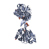
- Structure visualization
| Structure viewer | Molecule: MolmilJmol/JSmol |
|---|
- Downloads & links
-Download
| PDBx/mmCIF format | 1lyx.cif.gz | 66.1 KB | Display | PDBx/mmCIF format |
|---|---|---|---|---|
| PDB format | pdb1lyx.ent.gz | 48.7 KB | Display | PDB format |
| PDBx/mmJSON format | 1lyx.json.gz | Tree view | PDBx/mmJSON format | |
| Others |  Other downloads Other downloads |
-Validation report
| Arichive directory | https://data.pdbj.org/pub/pdb/validation_reports/ly/1lyxftp://data.pdbj.org/pub/pdb/validation_reports/ly/1lyx | HTTPS FTP |
|---|
-Related structure data
| Related structure data | 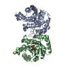 1lzoC 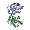 1ydvS C: citing same article ( S: Starting model for refinement |
|---|---|
| Similar structure data |


-Links
 PDBj
PDBj
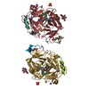
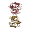
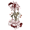
- Assembly
| Deposited unit | 
| ||||||||||
|---|---|---|---|---|---|---|---|---|---|---|---|
| 1 | 
| ||||||||||
| 2 | 
| ||||||||||
| Unit cell |
|
-Components
| #1: Protein | / E.C.5.3.1.1 / TIM / triose-phosphate isomerase / triose phosphate isomerase Mass: 27997.738 Da / Num. of mol.: 1 Source method: isolated from a genetically manipulated source Source: (gene. exp.) Plasmodium falciparum (malaria parasite P. falciparum)Gene: TPI / Plasmid: ptrc 99A vector, called pARC / Production host:  Escherichia coli (E. coli) / Strain (production host): AA200 / References: UniProt: Q07412, triose-phosphate isomerase Escherichia coli (E. coli) / Strain (production host): AA200 / References: UniProt: Q07412, triose-phosphate isomerase |
|---|---|
| #2: Chemical | ChemComp-PGA / 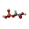  Mass: 156.031 Da / Num. of mol.: 1 Mass: 156.031 Da / Num. of mol.: 1Source method: isolated from a genetically manipulated source Formula: C2H5O6P |
| #3: Water | ChemComp-HOH / Water Mass: 18.015 Da / Num. of mol.: 173 / Source method: isolated from a natural source / Formula: H2O Mass: 18.015 Da / Num. of mol.: 173 / Source method: isolated from a natural source / Formula: H2O |
-Experimental details
-Experiment
| Experiment | Method: X-RAY DIFFRACTION / Number of used crystals: 1 |
|---|
- Sample preparation
| Crystal | Density Matthews: 2.4 Å3/Da / Density % sol: 46.74 % | |||||||||||||||||||||||||||||||||||||||||||||||||
|---|---|---|---|---|---|---|---|---|---|---|---|---|---|---|---|---|---|---|---|---|---|---|---|---|---|---|---|---|---|---|---|---|---|---|---|---|---|---|---|---|---|---|---|---|---|---|---|---|---|---|
| Crystal grow | Temperature: 295 K / Method: vapor diffusion, hanging drop / pH: 6.5 Details: 12% to 25% PEG 6000 or PEG 4000 or PEG 3350 or PEG 1450 in the presence of EDTA, DTT and Sodium azide in 100mM MES. pH 6.5, VAPOR DIFFUSION, HANGING DROP at 295K | |||||||||||||||||||||||||||||||||||||||||||||||||
| Crystal grow | *PLUS | |||||||||||||||||||||||||||||||||||||||||||||||||
| Components of the solutions | *PLUS
|
-Data collection
| Diffraction | Mean temperature: 295 K |
|---|---|
| Diffraction source | Source: ROTATING ANODE / Type: RIGAKU RU200 / Wavelength: 1.5418 Å |
| Detector | Type: MARRESEARCH / Detector: IMAGE PLATE / Date: Feb 10, 2000 / Details: Mirrors |
| Radiation | Monochromator: Mirrors / Protocol: SINGLE WAVELENGTH / Monochromatic (M) / Laue (L): M / Scattering type: x-ray |
| Radiation wavelength | Wavelength: 1.5418 Å / Relative weight: 1 |
| Reflection | Resolution: 1.9→20 Å / Num. all: 20508 / Num. obs: 20508 / % possible obs: 98.4 % / Redundancy: 18.8 % / Rsym value: 0.068 |
| Reflection shell | Resolution: 1.9→1.97 Å / Num. unique all: 2008 / Rsym value: 0.215 / % possible all: 97.1 |
| Reflection | *PLUS Highest resolution: 1.9 Å / Num. obs: 10508 / Num. measured all: 198026 / Rmerge(I) obs: 0.068 |
- Processing
| Software |
| ||||||||||||||||||||||||||||||||||||
|---|---|---|---|---|---|---|---|---|---|---|---|---|---|---|---|---|---|---|---|---|---|---|---|---|---|---|---|---|---|---|---|---|---|---|---|---|---|
| Refinement | Method to determine structure: MOLECULAR REPLACEMENT Starting model: Monomer of unbound PfTIM, PDB code 1YDV Resolution: 1.9→20 Å / Data cutoff high absF: 416213.98 / Data cutoff low absF: 0 / Isotropic thermal model: Isotropic / Cross valid method: THROUGHOUT / σ(F): 0.1 / Stereochemistry target values: Engh & Huber Details: Bulk solvent and anisotropic B-scaling were applied throughout the refinement
| ||||||||||||||||||||||||||||||||||||
| Solvent computation | Solvent model: FLAT MODEL / Bsol: 47.54 Å2 / ksol: 0.355873 e/Å3 | ||||||||||||||||||||||||||||||||||||
| Displacement parameters | Biso mean: 23.74 Å2
| ||||||||||||||||||||||||||||||||||||
| Refine analyze |
| ||||||||||||||||||||||||||||||||||||
| Refinement step | Cycle: LAST / Resolution: 1.9→20 Å
| ||||||||||||||||||||||||||||||||||||
| Refine LS restraints |
| ||||||||||||||||||||||||||||||||||||
| LS refinement shell | Resolution: 1.9→1.97 Å / Total num. of bins used: 10
| ||||||||||||||||||||||||||||||||||||
| Xplor file |
| ||||||||||||||||||||||||||||||||||||
| Refinement | *PLUS Highest resolution: 1.9 Å / Rfactor Rfree: 0.211 / Rfactor Rwork: 0.18 | ||||||||||||||||||||||||||||||||||||
| Solvent computation | *PLUS | ||||||||||||||||||||||||||||||||||||
| Displacement parameters | *PLUS | ||||||||||||||||||||||||||||||||||||
| Refine LS restraints | *PLUS
|
