 Movie
Movie Controller

[English] 日本語
 Yorodumi
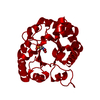
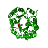
- PDB-1n55: 0.83A resolution structure of the E65Q mutant of Leishmania mexic... -
+ Open data

- Basic information
| Entry | Database: PDB / ID: 1n55 | ||||||
|---|---|---|---|---|---|---|---|
| Title | 0.83A resolution structure of the E65Q mutant of Leishmania mexicana triosephosphate isomerase complexed with 2-phosphoglycolate | ||||||
 Components Components | triosephosphate isomerase | ||||||
 Keywords Keywords | ISOMERASE / TIM / atomic resolution / enzyme-ligand complex / transition-state analogue / low-barrier hydrogen bond | ||||||
| Function / homology |  Function and homology informationglycosome / glyceraldehyde-3-phosphate biosynthetic process / glycerol catabolic process / triose-phosphate isomerase / triose-phosphate isomerase activity / gluconeogenesis / glycolytic process / cytosol Function and homology informationglycosome / glyceraldehyde-3-phosphate biosynthetic process / glycerol catabolic process / triose-phosphate isomerase / triose-phosphate isomerase activity / gluconeogenesis / glycolytic process / cytosolSimilarity search - Function | ||||||
| Biological species |   Leishmania mexicana (eukaryote) Leishmania mexicana (eukaryote) | ||||||
| Method | X-RAY DIFFRACTION / SYNCHROTRON / MOLECULAR REPLACEMENT / Resolution: 0.83 Å | ||||||
 Authors Authors | Kursula, I. / Wierenga, R.K. | ||||||
 Citation Citation | Journal: J.Biol.Chem. / Year: 2003 Title: Crystal structure of triosephosphate isomerase complexed with 2-phosphoglycolate at 0.83-A resolution Authors: Kursula, I. / Wierenga, R.K. | ||||||
| History |
|
- Structure visualization
| Structure viewer | Molecule: MolmilJmol/JSmol |
|---|
- Downloads & links
-Download
| PDBx/mmCIF format | 1n55.cif.gz | 140.9 KB | Display | PDBx/mmCIF format |
|---|---|---|---|---|
| PDB format | pdb1n55.ent.gz | 109.9 KB | Display | PDB format |
| PDBx/mmJSON format | 1n55.json.gz | Tree view | PDBx/mmJSON format | |
| Others |  Other downloads Other downloads |
-Validation report
| Arichive directory | https://data.pdbj.org/pub/pdb/validation_reports/n5/1n55ftp://data.pdbj.org/pub/pdb/validation_reports/n5/1n55 | HTTPS FTP |
|---|
-Related structure data
| Related structure data | |
|---|---|
| Similar structure data |


-Links
 PDBj
PDBj


- Assembly
| Deposited unit | 
| ||||||||||||
|---|---|---|---|---|---|---|---|---|---|---|---|---|---|
| 1 | 
| ||||||||||||
| Unit cell |
| ||||||||||||
| Components on special symmetry positions |
|
-Components
| #1: Protein | / TIM Mass: 27208.236 Da / Num. of mol.: 1 / Mutation: E65Q Source method: isolated from a genetically manipulated source Source: (gene. exp.) Leishmania mexicana (eukaryote) / Production host:  Escherichia coli (E. coli) / References: UniProt: P48499, triose-phosphate isomerase Escherichia coli (E. coli) / References: UniProt: P48499, triose-phosphate isomerase | ||||
|---|---|---|---|---|---|
| #2: Chemical | ChemComp-PGA / 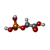  Mass: 156.031 Da / Num. of mol.: 1 Mass: 156.031 Da / Num. of mol.: 1Source method: isolated from a genetically manipulated source Formula: C2H5O6P | ||||
| #3: Chemical | Acetic acid  Mass: 60.052 Da / Num. of mol.: 2 / Source method: obtained synthetically / Formula: C2H4O2 Mass: 60.052 Da / Num. of mol.: 2 / Source method: obtained synthetically / Formula: C2H4O2#4: Chemical | Glycerol  Mass: 92.094 Da / Num. of mol.: 3 / Source method: obtained synthetically / Formula: C3H8O3 Mass: 92.094 Da / Num. of mol.: 3 / Source method: obtained synthetically / Formula: C3H8O3#5: Water | ChemComp-HOH / | Water Mass: 18.015 Da / Num. of mol.: 561 / Source method: isolated from a natural source / Formula: H2O Mass: 18.015 Da / Num. of mol.: 561 / Source method: isolated from a natural source / Formula: H2O |
-Experimental details
-Experiment
| Experiment | Method: X-RAY DIFFRACTION / Number of used crystals: 1 |
|---|
- Sample preparation
| Crystal | Density Matthews: 2.42 Å3/Da / Density % sol: 49.09 % | ||||||||||||||||||
|---|---|---|---|---|---|---|---|---|---|---|---|---|---|---|---|---|---|---|---|
| Crystal grow | Temperature: 295 K / Method: vapor diffusion, hanging drop / pH: 4.5 Details: 20% PEG 6000, 0.1 M Na-acetate/NaOH, pH 4.5, VAPOR DIFFUSION, HANGING DROP, temperature 295K | ||||||||||||||||||
| Crystal grow | *PLUS Details: Kursula, I., (2001) Eur. J. Biochem., 268, 5189. | ||||||||||||||||||
| Components of the solutions | *PLUS
|
-Data collection
| Diffraction | Mean temperature: 100 K |
|---|---|
| Diffraction source | Source: SYNCHROTRON / Site: EMBL/DESY, HAMBURG  / Beamline: BW7B / Wavelength: 0.84 / Beamline: BW7B / Wavelength: 0.84 |
| Detector | Type: MAR scanner 345 mm plate / Detector: IMAGE PLATE / Details: Bent mirror |
| Radiation | Monochromator: Triangular / Protocol: SINGLE WAVELENGTH / Monochromatic (M) / Laue (L): M / Scattering type: x-ray |
| Radiation wavelength | Wavelength: 0.84 Å / Relative weight: 1 |
| Reflection | Resolution: 0.83→25 Å / Num. all: 243382 / Num. obs: 241678 / % possible obs: 99.3 % / Redundancy: 4.3 % / Biso Wilson estimate: 6.88 Å2 / Rmerge(I) obs: 0.029 / Net I/σ(I): 34 |
| Reflection shell | Resolution: 0.83→0.84 Å / Rmerge(I) obs: 0.394 / Mean I/σ(I) obs: 3 / % possible all: 97.2 |
| Reflection | *PLUS Lowest resolution: 25 Å |
| Reflection shell | *PLUS % possible obs: 97.2 % / Num. unique obs: 11796 |
- Processing
| Software |
| ||||||||||||||||||
|---|---|---|---|---|---|---|---|---|---|---|---|---|---|---|---|---|---|---|---|
| Refinement | Method to determine structure: MOLECULAR REPLACEMENT / Resolution: 0.83→25 Å / Cross valid method: THROUGHOUT / σ(F): 0
| ||||||||||||||||||
| Refinement step | Cycle: LAST / Resolution: 0.83→25 Å
| ||||||||||||||||||
| Refine LS restraints |
| ||||||||||||||||||
| Software | *PLUS Name: SHELXL / Version: 97 / Classification: refinement | ||||||||||||||||||
| Refinement | *PLUS Lowest resolution: 25 Å / Rfactor obs: 0.1026 | ||||||||||||||||||
| Solvent computation | *PLUS | ||||||||||||||||||
| Displacement parameters | *PLUS | ||||||||||||||||||
| Refine LS restraints | *PLUS Type: s_plane_restr / Dev ideal: 0.441 |
