 Movie
Movie Controller

[English] 日本語
 Yorodumi
- PDB-4jeq: Different Contribution of Conserved Amino Acids to the Global Pro... -
+ Open data

- Basic information
| Entry | Database: PDB / ID: 4jeq | ||||||
|---|---|---|---|---|---|---|---|
| Title | Different Contribution of Conserved Amino Acids to the Global Properties of Homologous Enzymes | ||||||
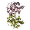
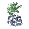
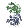
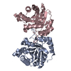
 Components Components | TRIOSEPHOSPHATE ISOMERASE, GLYCOSOMAL | ||||||
 Keywords Keywords | ISOMERASE / TIM BARREL / DISEASE MUTATION / PENTOSE SHUNT / GLUCONEOGENESIS / GLYCOLYSIS | ||||||
| Function / homology |  Function and homology informationglycosome / triose-phosphate isomerase / triose-phosphate isomerase activity / gluconeogenesis / glycolytic process / cytoplasm Function and homology informationglycosome / triose-phosphate isomerase / triose-phosphate isomerase activity / gluconeogenesis / glycolytic process / cytoplasmSimilarity search - Function | ||||||
| Biological species |  TRYPANOSOMA CRUZI (eukaryote) TRYPANOSOMA CRUZI (eukaryote) | ||||||
| Method | X-RAY DIFFRACTION / SYNCHROTRON / MOLECULAR REPLACEMENT / Resolution: 2.303 Å | ||||||
 Authors Authors | Hernandez-Santoyo, A. / Aguirre-Fuentes, Y. / Torres-Larios, A. / Gomez-Puyou, A. / De Gomez-Puyou, M.T. | ||||||
 Citation Citation | Journal: Proteins / Year: 2014 Title: Different contribution of conserved amino acids to the global properties of triosephosphate isomerases. Authors: Aguirre, Y. / Cabrera, N. / Aguirre, B. / Perez-Montfort, R. / Hernandez-Santoyo, A. / Reyes-Vivas, H. / Enriquez-Flores, S. / de Gomez-Puyou, M.T. / Gomez-Puyou, A. / Sanchez-Ruiz, J.M. / Costas, M. | ||||||
| History |
|
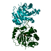
- Structure visualization
| Structure viewer | Molecule: MolmilJmol/JSmol |
|---|
- Downloads & links
-Download
| PDBx/mmCIF format | 4jeq.cif.gz | 555.8 KB | Display | PDBx/mmCIF format |
|---|---|---|---|---|
| PDB format | pdb4jeq.ent.gz | 462.3 KB | Display | PDB format |
| PDBx/mmJSON format | 4jeq.json.gz | Tree view | PDBx/mmJSON format | |
| Others |  Other downloads Other downloads |
-Validation report
| Arichive directory | https://data.pdbj.org/pub/pdb/validation_reports/je/4jeqftp://data.pdbj.org/pub/pdb/validation_reports/je/4jeq | HTTPS FTP |
|---|
-Related structure data
| Related structure data |  4hhpC 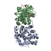 2j27S C: citing same article ( S: Starting model for refinement |
|---|---|
| Similar structure data |


-Links
 PDBj
PDBj
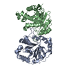
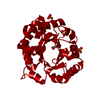
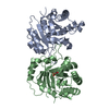
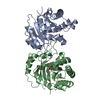
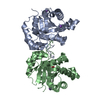
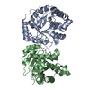
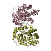
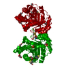
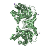
- Assembly
| Deposited unit | 
| ||||||||
|---|---|---|---|---|---|---|---|---|---|
| 1 | 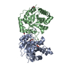
| ||||||||
| 2 | 
| ||||||||
| 3 | 
| ||||||||
| 4 | 
| ||||||||
| 5 | 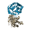
| ||||||||
| 6 | 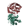
| ||||||||
| Unit cell |
|
-Components
| #1: Protein | / TIM / TRIOSE-PHOSPHATE ISOMERASE Mass: 26851.807 Da / Num. of mol.: 12 / Mutation: E104D Source method: isolated from a genetically manipulated source Source: (gene. exp.) TRYPANOSOMA CRUZI (eukaryote) / Strain: MEXICAN NINOA / Plasmid: PET3A / Production host:  ESCHERICHIA COLI (E. coli) / Strain (production host): BL21(DE3) / References: UniProt: P04789, triose-phosphate isomerase ESCHERICHIA COLI (E. coli) / Strain (production host): BL21(DE3) / References: UniProt: P04789, triose-phosphate isomerase#2: Chemical | ChemComp-SO4 / Sulfate  Mass: 96.063 Da / Num. of mol.: 5 / Source method: obtained synthetically / Formula: SO4 Mass: 96.063 Da / Num. of mol.: 5 / Source method: obtained synthetically / Formula: SO4#3: Chemical | ChemComp-PEG / Diethylene glycol  Mass: 106.120 Da / Num. of mol.: 5 / Source method: obtained synthetically / Formula: C4H10O3 Mass: 106.120 Da / Num. of mol.: 5 / Source method: obtained synthetically / Formula: C4H10O3#4: Water | ChemComp-HOH / | Water Mass: 18.015 Da / Num. of mol.: 594 / Source method: isolated from a natural source / Formula: H2O Mass: 18.015 Da / Num. of mol.: 594 / Source method: isolated from a natural source / Formula: H2O |
|---|
-Experimental details
-Experiment
| Experiment | Method: X-RAY DIFFRACTION / Number of used crystals: 1 |
|---|
- Sample preparation
| Crystal | Density Matthews: 2.18 Å3/Da / Density % sol: 43.69 % |
|---|---|
| Crystal grow | Temperature: 281.15 K / Method: vapor diffusion, sitting drop / pH: 8.6 Details: 25% w/v PEG monomethyl ether 2000, 0.1 M Tris, 0.01 M Nickel (II) chloride hexahydrate, 5% w/v n-dodecyl-N,N-dimethylamin-N-oxide , pH 8.6, VAPOR DIFFUSION, SITTING DROP, temperature 281.15K |
-Data collection
| Diffraction | Mean temperature: 113 K | ||||||||||||||||||
|---|---|---|---|---|---|---|---|---|---|---|---|---|---|---|---|---|---|---|---|
| Diffraction source | Source: SYNCHROTRON / Type: OTHER / Wavelength: 0.9791 Å | ||||||||||||||||||
| Detector | Type: MARRESEARCH / Detector: CCD / Date: Jul 28, 2010 Details: HIGH-RESOLUTION DOUBLE- CRYSTAL SI(220) SAGITTAL FOCUSING, ROSENBAUM-ROCK VERTICAL FOCUSING MIRROR | ||||||||||||||||||
| Radiation | Monochromator: ROSENBAUM-ROCK MONOCHROMATOR / Protocol: SINGLE WAVELENGTH / Monochromatic (M) / Laue (L): M / Scattering type: x-ray | ||||||||||||||||||
| Radiation wavelength | Wavelength: 0.9791 Å / Relative weight: 1 | ||||||||||||||||||
| Reflection | Resolution: 2.3→89.78 Å / Num. all: 122434 / Num. obs: 122434 / % possible obs: 99.6 % / Observed criterion σ(F): 0 / Observed criterion σ(I): 0 / Redundancy: 3 % / Biso Wilson estimate: 36.51 Å2 / Rmerge(I) obs: 0.089 / Rsym value: 0.086 / Net I/σ(I): 8.1 | ||||||||||||||||||
| Reflection shell |
|
- Processing
| Software |
| |||||||||||||||||||||||||||||||||||||||||||||||||||||||||||||||||||||||||||||||||||||||||||||||||||||||||||||||||||||||||||||||||||||||||||||||||||||||||||||||||||||||||||||||||||||||||||||||||||||||||||||||||||||||||
|---|---|---|---|---|---|---|---|---|---|---|---|---|---|---|---|---|---|---|---|---|---|---|---|---|---|---|---|---|---|---|---|---|---|---|---|---|---|---|---|---|---|---|---|---|---|---|---|---|---|---|---|---|---|---|---|---|---|---|---|---|---|---|---|---|---|---|---|---|---|---|---|---|---|---|---|---|---|---|---|---|---|---|---|---|---|---|---|---|---|---|---|---|---|---|---|---|---|---|---|---|---|---|---|---|---|---|---|---|---|---|---|---|---|---|---|---|---|---|---|---|---|---|---|---|---|---|---|---|---|---|---|---|---|---|---|---|---|---|---|---|---|---|---|---|---|---|---|---|---|---|---|---|---|---|---|---|---|---|---|---|---|---|---|---|---|---|---|---|---|---|---|---|---|---|---|---|---|---|---|---|---|---|---|---|---|---|---|---|---|---|---|---|---|---|---|---|---|---|---|---|---|---|---|---|---|---|---|---|---|---|---|---|---|---|---|---|---|---|
| Refinement | Method to determine structure: MOLECULAR REPLACEMENT Starting model: PDB ENTRY 2J27 Resolution: 2.303→61.577 Å / SU ML: 0.31 / σ(F): 1.34 / Phase error: 28.35 / Stereochemistry target values: ML
| |||||||||||||||||||||||||||||||||||||||||||||||||||||||||||||||||||||||||||||||||||||||||||||||||||||||||||||||||||||||||||||||||||||||||||||||||||||||||||||||||||||||||||||||||||||||||||||||||||||||||||||||||||||||||
| Solvent computation | Shrinkage radii: 0.9 Å / VDW probe radii: 1.11 Å / Solvent model: FLAT BULK SOLVENT MODEL | |||||||||||||||||||||||||||||||||||||||||||||||||||||||||||||||||||||||||||||||||||||||||||||||||||||||||||||||||||||||||||||||||||||||||||||||||||||||||||||||||||||||||||||||||||||||||||||||||||||||||||||||||||||||||
| Refinement step | Cycle: LAST / Resolution: 2.303→61.577 Å
| |||||||||||||||||||||||||||||||||||||||||||||||||||||||||||||||||||||||||||||||||||||||||||||||||||||||||||||||||||||||||||||||||||||||||||||||||||||||||||||||||||||||||||||||||||||||||||||||||||||||||||||||||||||||||
| Refine LS restraints |
| |||||||||||||||||||||||||||||||||||||||||||||||||||||||||||||||||||||||||||||||||||||||||||||||||||||||||||||||||||||||||||||||||||||||||||||||||||||||||||||||||||||||||||||||||||||||||||||||||||||||||||||||||||||||||
| LS refinement shell |
|
