 ムービー
ムービー コントローラー
 構造ビューア
 EMN検索について


-検索条件
-検索結果
検索 (著者・登録者: carter & ap)の結果119件中、1から50件目までを表示しています

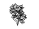
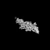
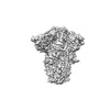
EMDB-17835: 
Consensus cryo-EM structure of Dynein-Dynactin-JIP3(1-185)-LIS1

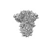
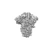
EMDB-17836:
Consensus cryo-EM structure of Dynein-dynactin-JIP3(1-560)-LIS1

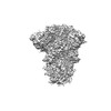
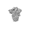
EMDB-17825:
Cytoplasmic dynein-1 motor domain in post-powerstroke state

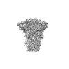
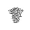
EMDB-17826:
Cytoplasmic dynein-1 motor domain bound to dynactin-p150glued and LIS1
EMDB-17828:
Cytoplasmic dynein-1 motor domain bound to LIS1

EMDB-17829:
Cytoplasmic dynein-1 A1/A2 motor domains bound to LIS1

EMDB-17830:
Cytoplasmic dynein-A heavy chain bound to dynactin-p150glued and IC-LC tower

EMDB-17831:
Cytoplasmic dynein-B heavy chain bound to IC-LC tower

EMDB-17832:
Cytoplasmic dynein-1 heavy chain bound to JIP3-LZI

EMDB-17833:
Cytoplasmic dynein-1 heavy chain bound to JIP3-RH1

EMDB-17834:
Dynactin pointed end bound to JIP3

EMDB-17873:
Composite structure of Dynein-Dynactin-JIP3-LIS1

EMDB-18121:
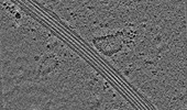
A cryo-ET study of ciliary rootlet organization - Example cell-like tomogram

EMDB-18122:
A cryo-ET study of ciliary rootlet organization - purified rootlet example 1

EMDB-18123:
A cryo-ET study of ciliary rootlet organization - purified rootlet example 2

EMDB-18124:
A cryo-ET study of ciliary rootlet organization - purified rootlet example 3

EMDB-18125:
A cryo-ET study of ciliary rootlet organization - purified rootlet example 4
EMDB-18475:
Cryo-electron tomogram of an induced S2 cell protrusion. The cell was treated with 2uM thapsigargin (5h) and with 2.5uM Cytochalasin D (2h).
EMDB-16877:
Subtomogram averaging structure of cofilactin filament inside microtubule lumen of Drosophila S2 cell protrusion.
EMDB-16685:
Tomogram of an induced protrusion of a Drosophila S2 cell
EMDB-16693:
Tomogram of an induced protrusion of a Drosophila S2 cell
EMDB-16695:
Tomogram of an induced protrusion of a Drosophila S2 alpha-tubulin acetyltransferase knock-out (dTAT KO) cell with a filament inside the microtubule lumen
EMDB-16720:
Tomogram of an induced protrusion of a Drosophila S2 cell with filaments inside the microtubule lumen.
EMDB-16800:
Tomogram of an induced protrusion of a cofilin knock-down Drosophila S2 cell with filaments inside the microtubule lumen
EMDB-16811:
Tomogram of an induced protrusion of a cofilin knock-down Drosophila S2 cell with a filament inside the microtubule lumen.
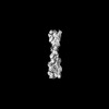
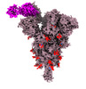
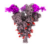
EMDB-33748:
Spike_GSAS_6P protomer RBD domain bound with R1-32 Fab and ACE2 with 3:3:3 ratio
EMDB-33760:
Spike_GSAS_6P and R1-32 Fab with 3to1 ratio
EMDB-33764:
SARS-COV-2 Spike_GSAS_6P bound with two R1-32 Fabs
EMDB-33766:
SARS-CoV-2 Spike (6P) in complex with 3 R1-32 Fabs

EMDB-33772:
SARS-CoV-2 Spike (6P) in complex with 3 R1-32 Fabs and 3 ACE2
EMDB-14552:
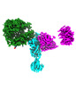
The barbed end complex of dynactin bound to BICDR1 and the cytoplasmic dynein tails (A2, B1, B2)
EMDB-14549:
Composite structure of dynein-dynactin-BICDR on microtubules

EMDB-14550:
Cytoplasmic dynein-1 motor domain
EMDB-14551:
Cytoplasmic dynein-1 motor domain AAA1, AAA2, and AAA3 subunits
EMDB-14553:
Cytoplasmic dynein (A2) bound to BICDR1
EMDB-14555:
Cytoplasmic dynein (A1) bound to BICDR1
EMDB-14556:
Cytoplasmic dynein light intermediate chain (B1) bound to the motor domain (A2).
EMDB-14559:
The pointed end complex of dynactin bound to BICDR1
EMDB-15396:
Consensus map of dynein-dynactin-BICDR on microtubules
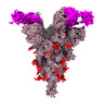
EMDB-33453:
Structure of SARS-CoV-2 Spike Protein with Engineered x3 Disulfide (x3(D427C, V987C) and single Arg S1/S2 cleavage site), Locked-1 Conformation
EMDB-33454:
Structure of SARS-CoV-2 Spike Protein with Engineered x3 Disulfide (x3(D427C, V987C) and single Arg S1/S2 cleavage site), Locked-211 Conformation
EMDB-33455:
Structure of SARS-CoV-2 Spike Protein with Engineered x3 Disulfide (x3(D427C, V987C) and single Arg S1/S2 cleavage site), Locked-122 Conformation
EMDB-33456:
Structure of SARS-CoV-2 Spike Protein with Engineered x3 Disulfide (x3(D427C, V987C) and single Arg S1/S2 cleavage site), Locked-2 Conformation
EMDB-33457:
Structure of SARS-CoV-2 Spike Protein with Engineered x3 Disulfide (x3(D427C, V987C) and single Arg S1/S2 cleavage site), Closed Conformation
EMDB-33458:
Structure of SARS-CoV-2 D614G Spike Protein with Engineered x3 Disulfide (x3(D427C, V987C) and single Arg S1/S2 cleavage site), Locked-2 Conformation
EMDB-33459:
Structure of SARS-CoV-2 D614G Spike Protein with Engineered x3 Disulfide (x3(D427C, V987C) and single Arg S1/S2 cleavage site), Closed Conformation
EMDB-33460:
Structure of SARS-CoV-2 Spike Protein with Engineered x3 Disulfide (x3(D427C, V987C) and single Arg S1/S2 cleavage site), incubated in Low pH after 40-Day Storage in PBS, Locked-2 Conformation
EMDB-33461:
Structure of SARS-CoV-2 Spike Protein with Engineered x3 Disulfide (x3(D427C, V987C) and single Arg S1/S2 cleavage site) after 40-Day Storage in PBS, Closed Conformation
EMDB-33462:
Structure of SARS-CoV-2 Spike Protein with Engineered x3 Disulfide (x3(D427C, V987C) and single Arg S1/S2 cleavage site), incubated in Low pH after 40-Day Storage in PBS, Closed Conformation
EMDB-33463:
Structure of SARS-CoV-2 D614G spike protein (single Arg S1/S2 cleavage site), Closed Conformation
ページ:
 wwPDBはEMDBデータモデルのバージョン3へ移行します
