Movie
Movie Controller
 Structure viewers
 About Yorodumi Papers


+Search query
-Structure paper
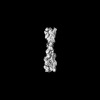
| Title | CryoET shows cofilactin filaments inside the microtubule lumen. |
|---|---|
| Journal, issue, pages | bioRxiv, Year 2023 |
| Publish date | Mar 31, 2023 |
 Authors Authors | Camilla Ventura Santos / Stephen L Rogers / Andrew P Carter /  |
| PubMed Abstract | Cytoplasmic microtubules are tubular polymers that can harbor small proteins or filaments inside their lumen. The identity of these objects and what causes their accumulation has not been ...Cytoplasmic microtubules are tubular polymers that can harbor small proteins or filaments inside their lumen. The identity of these objects and what causes their accumulation has not been conclusively established. Here, we used cryogenic electron tomography (cryoET) of S2 cell protrusions and found filaments inside the microtubule lumen, which resemble those reported recently in human HAP1 cells. The frequency of these filaments increased upon inhibition of the sarco/endoplasmic reticulum Ca ATPase (SERCA) with the small-molecule drug thapsigargin. Subtomogram averaging showed that the luminal filaments adopt a helical structure reminiscent of cofilin-bound actin (cofilactin). Consistent with this, cofilin was activated in cells under the same conditions that increased luminal filament occurrence. Furthermore, RNAi knock-down of cofilin reduced the frequency of luminal filaments with cofilactin morphology. These results suggest that cofilin activation stimulates its accumulation on actin filaments inside the microtubule lumen. |
 External links External links | bioRxiv / PubMed:37034688 / PubMed Central |
| Methods | EM (tomography) / EM (subtomogram averaging) |
| Resolution | 16.5 Å |
| Structure data |  EMDB-16685: Tomogram of an induced protrusion of a Drosophila S2 cell  EMDB-16693: Tomogram of an induced protrusion of a Drosophila S2 cell  EMDB-16695: Tomogram of an induced protrusion of a Drosophila S2 alpha-tubulin acetyltransferase knock-out (dTAT KO) cell with a filament inside the microtubule lumen  EMDB-16720: Tomogram of an induced protrusion of a Drosophila S2 cell with filaments inside the microtubule lumen.  EMDB-16800: Tomogram of an induced protrusion of a cofilin knock-down Drosophila S2 cell with filaments inside the microtubule lumen  EMDB-16811: Tomogram of an induced protrusion of a cofilin knock-down Drosophila S2 cell with a filament inside the microtubule lumen. EMDB-16877, PDB-8oh4: |
| Source |
|
 Keywords Keywords |  CONTRACTILE PROTEIN / Cytoskeleton / Filament / Actin / Cofilactin CONTRACTILE PROTEIN / Cytoskeleton / Filament / Actin / Cofilactin |