Loading
PDBj
MenuPDBj@FacebookPDBj@TwitterPDBj@YouTubewwPDB FoundationwwPDB
RCSB PDBPDBeBMRBAdv. SearchSearch help
Search by PDB author
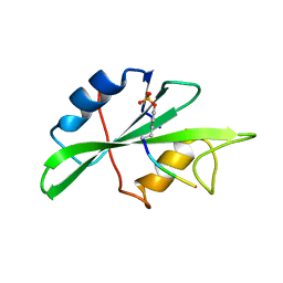
1AB2
DownloadVisualize
BU of 1ab2 by Molmil
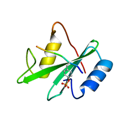
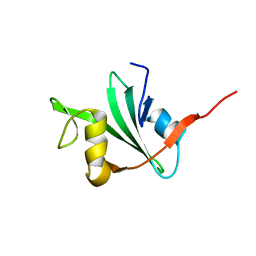
THREE-DIMENSIONAL SOLUTION STRUCTURE OF THE SRC HOMOLOGY 2 DOMAIN OF C-ABL
Descriptor: C-ABL TYROSINE KINASE SH2 DOMAIN
Authors:Overduin, M, Rios, C.B, Mayer, B.J, Baltimore, D, Cowburn, D.
Deposit date:1993-07-19
Release date:1994-01-31
Last modified:2022-02-16
Method:SOLUTION NMR
Cite:Three-dimensional solution structure of the src homology 2 domain of c-abl.
Cell(Cambridge,Mass.), 70, 1992
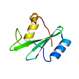
1AWO
DownloadVisualize
BU of 1awo by Molmil
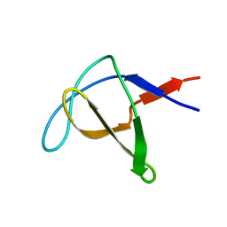
THE SOLUTION NMR STRUCTURE OF ABL SH3 AND ITS RELATIONSHIP TO SH2 IN THE SH(32) CONSTRUCT, 20 STRUCTURES
Descriptor: ABL TYROSINE KINASE
Authors:Cowburn, D.
Deposit date:1997-10-03
Release date:1998-01-28
Last modified:2022-02-16
Method:SOLUTION NMR
Cite:The solution structure of Abl SH3, and its relationship to SH2 in the SH(32) construct.
Structure, 3, 1995
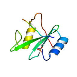
1AYC
DownloadVisualize
BU of 1ayc by Molmil
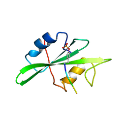
CRYSTAL STRUCTURES OF PEPTIDE COMPLEXES OF THE AMINO-TERMINAL SH2 DOMAIN OF THE SYP TYROSINE PHOSPHATASE
Descriptor: PEPTIDE PDGFR-740, PROTEIN-TYROSINE PHOSPHATASE SYP (N-TERMINAL SH2 DOMAIN)
Authors:Lee, C.-H, Kuriyan, J.
Deposit date:1994-05-15
Release date:1994-08-31
Last modified:2011-07-13
Method:X-RAY DIFFRACTION (2.3 Å)
Cite:Crystal structures of peptide complexes of the amino-terminal SH2 domain of the Syp tyrosine phosphatase.
Structure, 2, 1994
1AYA
DownloadVisualize
BU of 1aya by Molmil
CRYSTAL STRUCTURES OF PEPTIDE COMPLEXES OF THE AMINO-TERMINAL SH2 DOMAIN OF THE SYP TYROSINE PHOSPHATASE
Descriptor: PEPTIDE PDGFR-1009, PROTEIN-TYROSINE PHOSPHATASE SYP (N-TERMINAL SH2 DOMAIN)
Authors:Lee, C.-H, Kuriyan, J.
Deposit date:1994-05-15
Release date:1994-08-31
Last modified:2011-07-13
Method:X-RAY DIFFRACTION (2.05 Å)
Cite:Crystal structures of peptide complexes of the amino-terminal SH2 domain of the Syp tyrosine phosphatase.
Structure, 2, 1994
1SHB
DownloadVisualize
BU of 1shb by Molmil
CRYSTAL STRUCTURE OF THE PHOSPHOTYROSINE RECOGNITION DOMAIN SH2 OF V-SRC COMPLEXED WITH TYROSINE-PHOSPHORYLATED PEPTIDES
Descriptor: PHOSPHOPEPTIDE B, V-SRC SH2 DOMAIN
Authors:Waksman, G, Kuriyan, J.
Deposit date:1992-08-18
Release date:1993-10-31
Last modified:2011-07-13
Method:X-RAY DIFFRACTION (2 Å)
Cite:Crystal structure of the phosphotyrosine recognition domain SH2 of v-src complexed with tyrosine-phosphorylated peptides.
Nature, 358, 1992
1SHA
DownloadVisualize
BU of 1sha by Molmil
CRYSTAL STRUCTURE OF THE PHOSPHOTYROSINE RECOGNITION DOMAIN SH2 OF V-SRC COMPLEXED WITH TYROSINE-PHOSPHORYLATED PEPTIDES
Descriptor: PHOSPHOPEPTIDE A, V-SRC SH2 DOMAIN
Authors:Waksman, G, Kuriyan, J.
Deposit date:1992-08-18
Release date:1993-10-31
Last modified:2011-07-13
Method:X-RAY DIFFRACTION (1.5 Å)
Cite:Crystal structure of the phosphotyrosine recognition domain SH2 of v-src complexed with tyrosine-phosphorylated peptides.
Nature, 358, 1992
1AYB
DownloadVisualize
BU of 1ayb by Molmil
CRYSTAL STRUCTURES OF PEPTIDE COMPLEXES OF THE AMINO-TERMINAL SH2 DOMAIN OF THE SYP TYROSINE PHOSPHATASE
Descriptor: PEPTIDE IRS-1-895, PROTEIN-TYROSINE PHOSPHATASE SYP (N-TERMINAL SH2 DOMAIN)
Authors:Lee, C.-H, Kuriyan, J.
Deposit date:1994-05-15
Release date:1994-08-31
Last modified:2011-07-13
Method:X-RAY DIFFRACTION (3 Å)
Cite:Crystal structures of peptide complexes of the amino-terminal SH2 domain of the Syp tyrosine phosphatase.
Structure, 2, 1994
1AYD
DownloadVisualize
BU of 1ayd by Molmil
CRYSTAL STRUCTURES OF PEPTIDE COMPLEXES OF THE AMINO-TERMINAL SH2 DOMAIN OF THE SYP TYROSINE PHOSPHATASE
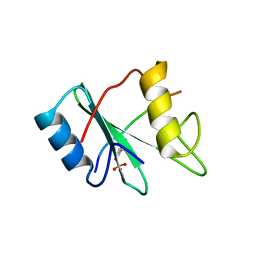
Descriptor: PROTEIN-TYROSINE PHOSPHATASE SYP (N-TERMINAL SH2 DOMAIN)
Authors:Lee, C.-H, Kuriyan, J.
Deposit date:1994-05-15
Release date:1994-08-31
Last modified:2024-02-07
Method:X-RAY DIFFRACTION (2.2 Å)
Cite:Crystal structures of peptide complexes of the amino-terminal SH2 domain of the Syp tyrosine phosphatase.
Structure, 2, 1994
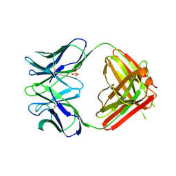
1NGQ
DownloadVisualize
BU of 1ngq by Molmil
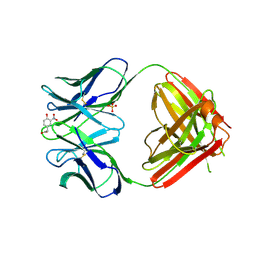
N1G9 (IGG1-LAMBDA) FAB FRAGMENT
Descriptor: N1G9 (IGG1-LAMBDA), SULFATE ION
Authors:Mizutani, R, Satow, Y.
Deposit date:1995-06-23
Release date:1996-07-11
Last modified:2018-04-04
Method:X-RAY DIFFRACTION (2.4 Å)
Cite:Three-dimensional structures of the Fab fragment of murine N1G9 antibody from the primary immune response and of its complex with (4-hydroxy-3-nitrophenyl)acetate.
J.Mol.Biol., 254, 1995
1NGP
DownloadVisualize
BU of 1ngp by Molmil
N1G9 (IGG1-LAMBDA) FAB FRAGMENT COMPLEXED WITH (4-HYDROXY-3-NITROPHENYL) ACETATE
Descriptor: 2-(4-HYDROXY-3-NITROPHENYL)ACETIC ACID, N1G9 (IGG1-LAMBDA), SULFATE ION
Authors:Mizutani, R, Satow, Y.
Deposit date:1995-06-23
Release date:1996-07-11
Last modified:2018-04-04
Method:X-RAY DIFFRACTION (2.4 Å)
Cite:Three-dimensional structures of the Fab fragment of murine N1G9 antibody from the primary immune response and of its complex with (4-hydroxy-3-nitrophenyl)acetate.
J.Mol.Biol., 254, 1995
1ABO
DownloadVisualize
BU of 1abo by Molmil
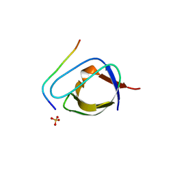
CRYSTAL STRUCTURE OF THE COMPLEX OF THE ABL TYROSINE KINASE SH3 DOMAIN WITH 3BP-1 SYNTHETIC PEPTIDE
Descriptor: 3BP-1 SYNTHETIC PEPTIDE, 10 RESIDUES, ABL TYROSINE KINASE, ...
Authors:Musacchio, A, Wilmanns, M, Saraste, M.
Deposit date:1995-05-19
Release date:1995-10-15
Last modified:2024-02-07
Method:X-RAY DIFFRACTION (2 Å)
Cite:High-resolution crystal structures of tyrosine kinase SH3 domains complexed with proline-rich peptides.
Nat.Struct.Biol., 1, 1994
1ABQ
DownloadVisualize
BU of 1abq by Molmil
CRYSTAL STRUCTURE OF THE UNLIGANDED ABL TYROSINE KINASE SH3 DOMAIN
Descriptor: ABL TYROSINE KINASE SRC-HOMOLOGY 3 (SH3) DOMAIN
Authors:Musacchio, A, Wilmanns, M, Saraste, M.
Deposit date:1995-05-19
Release date:1995-10-15
Last modified:2024-02-07
Method:X-RAY DIFFRACTION (2.8 Å)
Cite:High-resolution crystal structures of tyrosine kinase SH3 domains complexed with proline-rich peptides.
Nat.Struct.Biol., 1, 1994
1RDR
DownloadVisualize
BU of 1rdr by Molmil
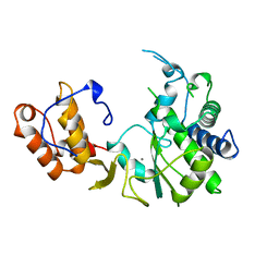
POLIOVIRUS 3D POLYMERASE
Descriptor: CALCIUM ION, POLIOVIRUS 3D POLYMERASE
Authors:Hansen, J, Long, A, Schultz, S.
Deposit date:1998-04-28
Release date:1998-09-16
Last modified:2024-02-14
Method:X-RAY DIFFRACTION (2.4 Å)
Cite:Structure of the RNA-dependent RNA polymerase of poliovirus.
Structure, 5, 1997

218853

PDB entries from 2024-04-24

PDB statisticsPDBj update infoContact PDBjnumon