 Movie
Movie Controller

[English] 日本語
 Yorodumi
- PDB-6zj6: Structure of the GH99 endo-alpha-mannanase from Bacteroides xylan... -
+ Open data

- Basic information
| Entry | Database: PDB / ID: 6zj6 | ||||||||||||||||||
|---|---|---|---|---|---|---|---|---|---|---|---|---|---|---|---|---|---|---|---|
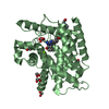
| Title | Structure of the GH99 endo-alpha-mannanase from Bacteroides xylanisolvens in complex with cyclohexylmethyl-Glc-1,3-isofagomine | ||||||||||||||||||
 Components Components | Glycosyl hydrolase family 71 | ||||||||||||||||||
 Keywords Keywords |  HYDROLASE / mannosidase / retaining HYDROLASE / mannosidase / retaining | ||||||||||||||||||
| Function / homology | Glycosyl hydrolase family 99 / Glycosyl hydrolase family 99 / hydrolase activity, acting on glycosyl bonds / membrane / ACETATE ION / alpha-D-glucopyranose / 5-HYDROXYMETHYL-3,4-DIHYDROXYPIPERIDINE / methylcyclohexane / Glycosyl hydrolase family 71 Function and homology information Function and homology information | ||||||||||||||||||
| Biological species |  Bacteroides xylanisolvens XB1A (bacteria) Bacteroides xylanisolvens XB1A (bacteria) | ||||||||||||||||||
| Method | X-RAY DIFFRACTION / SYNCHROTRON / MOLECULAR REPLACEMENT / Resolution: 1.09 Å | ||||||||||||||||||
 Authors Authors | Thompson, A.J. / Sobala, L.F. / Fernandes, P.Z. / Hakki, Z. / Howe, J.D. / Hill, M. / Zitzmann, N. / Davies, S. / Stamataki, Z. / Butters, T.D. ...Thompson, A.J. / Sobala, L.F. / Fernandes, P.Z. / Hakki, Z. / Howe, J.D. / Hill, M. / Zitzmann, N. / Davies, S. / Stamataki, Z. / Butters, T.D. / Alonzi, D.S. / Williams, S.J. / Davies, G.J. | ||||||||||||||||||
| Funding support |  United Kingdom, United Kingdom,  Australia, 5items Australia, 5items
| ||||||||||||||||||
 Citation Citation | Journal: Proc.Natl.Acad.Sci.USA / Year: 2020 Title: Structure of human endo-alpha-1,2-mannosidase (MANEA), an antiviral host-glycosylation target. Authors: Sobala, L.F. / Fernandes, P.Z. / Hakki, Z. / Thompson, A.J. / Howe, J.D. / Hill, M. / Zitzmann, N. / Davies, S. / Stamataki, Z. / Butters, T.D. / Alonzi, D.S. / Williams, S.J. / Davies, G.J. | ||||||||||||||||||
| History |
|
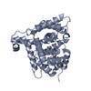
- Structure visualization
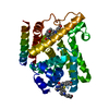
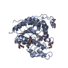
| Structure viewer | Molecule: MolmilJmol/JSmol |
|---|
- Downloads & links
-Download
| PDBx/mmCIF format | 6zj6.cif.gz | 184.2 KB | Display | PDBx/mmCIF format |
|---|---|---|---|---|
| PDB format | pdb6zj6.ent.gz | Display | PDB format | |
| PDBx/mmJSON format | 6zj6.json.gz | Tree view | PDBx/mmJSON format | |
| Others |  Other downloads Other downloads |
-Validation report
| Arichive directory | https://data.pdbj.org/pub/pdb/validation_reports/zj/6zj6ftp://data.pdbj.org/pub/pdb/validation_reports/zj/6zj6 | HTTPS FTP |
|---|
-Related structure data
| Related structure data |  6zdcC  6zdfC 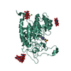 6zdkC 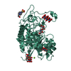 6zdlC 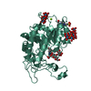 6zfaC 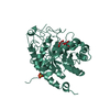 6zfnC  6zfqC  6zj1C 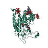 6zj5C  6hmgS C: citing same article ( S: Starting model for refinement |
|---|---|
| Similar structure data | |
| Experimental dataset #1 | Data reference: 10.5281/zenodo.4288341 / Data set type: diffraction image data |


-Links
 PDBj
PDBj

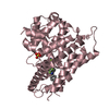
- Assembly
| Deposited unit | 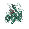
| ||||||||
|---|---|---|---|---|---|---|---|---|---|
| 1 |
| ||||||||
| Unit cell |
| ||||||||
| Components on special symmetry positions |
|
-Components
-Protein / Sugars , 2 types, 2 molecules AAA
| #1: Protein | Mass: 43933.594 Da / Num. of mol.: 1 Source method: isolated from a genetically manipulated source Source: (gene. exp.) Bacteroides xylanisolvens XB1A (bacteria)Gene: BXY_34140 / Production host: Escherichia coli BL21(DE3) (bacteria) / References: UniProt: D6D1V7 |
|---|---|
| #6: Sugar | ChemComp-GLC / Glucose Type: D-saccharide, alpha linking / Mass: 180.156 Da / Num. of mol.: 1 Type: D-saccharide, alpha linking / Mass: 180.156 Da / Num. of mol.: 1Source method: isolated from a genetically manipulated source Formula: C6H12O6 / Feature type: SUBJECT OF INVESTIGATION |
-Non-polymers , 5 types, 470 molecules 


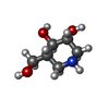

| #2: Chemical | ChemComp-EDO / Ethylene glycol Mass: 62.068 Da / Num. of mol.: 4 / Source method: obtained synthetically / Formula: C2H6O2 Mass: 62.068 Da / Num. of mol.: 4 / Source method: obtained synthetically / Formula: C2H6O2#3: Chemical | ChemComp-ACT / | Acetate Mass: 59.044 Da / Num. of mol.: 1 / Source method: obtained synthetically / Formula: C2H3O2 Mass: 59.044 Da / Num. of mol.: 1 / Source method: obtained synthetically / Formula: C2H3O2#4: Chemical | ChemComp-QTE / | Methylcyclohexane Mass: 98.186 Da / Num. of mol.: 1 / Source method: obtained synthetically / Formula: C7H14 / Feature type: SUBJECT OF INVESTIGATION Mass: 98.186 Da / Num. of mol.: 1 / Source method: obtained synthetically / Formula: C7H14 / Feature type: SUBJECT OF INVESTIGATION#5: Chemical | ChemComp-IFM / | Afegostat Mass: 147.172 Da / Num. of mol.: 1 / Source method: obtained synthetically / Formula: C6H13NO3 / Feature type: SUBJECT OF INVESTIGATION Mass: 147.172 Da / Num. of mol.: 1 / Source method: obtained synthetically / Formula: C6H13NO3 / Feature type: SUBJECT OF INVESTIGATION#7: Water | ChemComp-HOH / | WaterMass: 18.015 Da / Num. of mol.: 463 / Source method: isolated from a natural source / Formula: H2O |
|---|
-Details
| Has ligand of interest | Y |
|---|
-Experimental details
-Experiment
| Experiment | Method: X-RAY DIFFRACTION / Number of used crystals: 1 |
|---|
- Sample preparation
| Crystal | Density Matthews: 2.23 Å3/Da / Density % sol: 45 % |
|---|---|
| Crystal grow | Temperature: 292 K / Method: vapor diffusion, hanging drop / Details: 3 M sodium acetate pH 6.6 - 7,4 / PH range: 6.6-7.4 |
-Data collection
| Diffraction | Mean temperature: 100 K / Serial crystal experiment: N |
|---|---|
| Diffraction source | Source: SYNCHROTRON / Site: Diamond / Beamline: I03 / Wavelength: 0.9763 Å |
| Detector | Type: DECTRIS PILATUS 6M-F / Detector: PIXEL / Date: Dec 12, 2011 |
| Radiation | Protocol: SINGLE WAVELENGTH / Monochromatic (M) / Laue (L): M / Scattering type: x-ray |
| Radiation wavelength | Wavelength: 0.9763 Å / Relative weight: 1 |
| Reflection | Resolution: 1.09→76.35 Å / Num. obs: 153951 / % possible obs: 95.9 % / Redundancy: 5.1 % / Biso Wilson estimate: 11.6 Å2 / CC1/2: 0.997 / Rmerge(I) obs: 0.059 / Rpim(I) all: 0.027 / Rrim(I) all: 0.065 / Χ2: 0.78 / Net I/σ(I): 12.5 |
| Reflection shell | Resolution: 1.09→1.11 Å / Redundancy: 2.8 % / Rmerge(I) obs: 1.12 / Mean I/σ(I) obs: 0.9 / Num. unique obs: 5269 / CC1/2: 0.358 / Rpim(I) all: 0.715 / Rrim(I) all: 1.342 / Χ2: 0.43 / % possible all: 66.4 |
- Processing
| Software |
| |||||||||||||||||||||||||||||||||||||||||||||||||||||||||||||||||||||||||||||||||||||||||||||||||||||||||||||||||||||||||||||||||||||||||||||||||||||||||||||||||||||||||||||||||||||||||||||||||||||||||||||||||||||||||||||||||||||||
|---|---|---|---|---|---|---|---|---|---|---|---|---|---|---|---|---|---|---|---|---|---|---|---|---|---|---|---|---|---|---|---|---|---|---|---|---|---|---|---|---|---|---|---|---|---|---|---|---|---|---|---|---|---|---|---|---|---|---|---|---|---|---|---|---|---|---|---|---|---|---|---|---|---|---|---|---|---|---|---|---|---|---|---|---|---|---|---|---|---|---|---|---|---|---|---|---|---|---|---|---|---|---|---|---|---|---|---|---|---|---|---|---|---|---|---|---|---|---|---|---|---|---|---|---|---|---|---|---|---|---|---|---|---|---|---|---|---|---|---|---|---|---|---|---|---|---|---|---|---|---|---|---|---|---|---|---|---|---|---|---|---|---|---|---|---|---|---|---|---|---|---|---|---|---|---|---|---|---|---|---|---|---|---|---|---|---|---|---|---|---|---|---|---|---|---|---|---|---|---|---|---|---|---|---|---|---|---|---|---|---|---|---|---|---|---|---|---|---|---|---|---|---|---|---|---|---|---|---|---|---|---|---|
| Refinement | Method to determine structure: MOLECULAR REPLACEMENT Starting model: 6HMG Resolution: 1.09→76.35 Å / Cor.coef. Fo:Fc: 0.986 / Cor.coef. Fo:Fc free: 0.983 / SU B: 0.839 / SU ML: 0.017 / Cross valid method: FREE R-VALUE / ESU R: 0.023 / ESU R Free: 0.024 Details: Hydrogens have been added in their riding positions
| |||||||||||||||||||||||||||||||||||||||||||||||||||||||||||||||||||||||||||||||||||||||||||||||||||||||||||||||||||||||||||||||||||||||||||||||||||||||||||||||||||||||||||||||||||||||||||||||||||||||||||||||||||||||||||||||||||||||
| Solvent computation | Ion probe radii: 0.8 Å / Shrinkage radii: 0.8 Å / VDW probe radii: 1.2 Å / Solvent model: MASK BULK SOLVENT | |||||||||||||||||||||||||||||||||||||||||||||||||||||||||||||||||||||||||||||||||||||||||||||||||||||||||||||||||||||||||||||||||||||||||||||||||||||||||||||||||||||||||||||||||||||||||||||||||||||||||||||||||||||||||||||||||||||||
| Displacement parameters | Biso mean: 17.312 Å2
| |||||||||||||||||||||||||||||||||||||||||||||||||||||||||||||||||||||||||||||||||||||||||||||||||||||||||||||||||||||||||||||||||||||||||||||||||||||||||||||||||||||||||||||||||||||||||||||||||||||||||||||||||||||||||||||||||||||||
| Refinement step | Cycle: LAST / Resolution: 1.09→76.35 Å
| |||||||||||||||||||||||||||||||||||||||||||||||||||||||||||||||||||||||||||||||||||||||||||||||||||||||||||||||||||||||||||||||||||||||||||||||||||||||||||||||||||||||||||||||||||||||||||||||||||||||||||||||||||||||||||||||||||||||
| Refine LS restraints |
| |||||||||||||||||||||||||||||||||||||||||||||||||||||||||||||||||||||||||||||||||||||||||||||||||||||||||||||||||||||||||||||||||||||||||||||||||||||||||||||||||||||||||||||||||||||||||||||||||||||||||||||||||||||||||||||||||||||||
| LS refinement shell | Refine-ID: X-RAY DIFFRACTION / Total num. of bins used: 20
|
