 Movie
Movie Controller

[English] 日本語
 Yorodumi
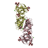
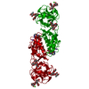
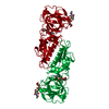
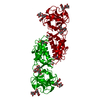
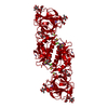
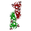
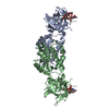
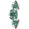
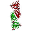
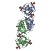
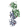
- PDB-6yh0: Marasmius oreades agglutinin (MOA) in complex with the truncated ... -
+ Open data

- Basic information
| Entry | Database: PDB / ID: 6yh0 | ||||||
|---|---|---|---|---|---|---|---|
| Title | Marasmius oreades agglutinin (MOA) in complex with the truncated PVPRAHS synthetic substrate | ||||||
 Components Components |
| ||||||
 Keywords Keywords |  TOXIN / fungal chimerolectin / papain-like cysteine protease / protease-substrate complex / calcium-binding protein / manganese-binding protein TOXIN / fungal chimerolectin / papain-like cysteine protease / protease-substrate complex / calcium-binding protein / manganese-binding protein | ||||||
| Function / homology | Agglutinin, C-terminal / Agglutinin C-terminal / Ricin-type beta-trefoil lectin domain-like / Lectin domain of ricin B chain profile. / Ricin B, lectin domain / Ricin B-like lectins / Papain-like cysteine peptidase superfamily / metal ion binding / Agglutinin Function and homology information Function and homology information | ||||||
| Biological species |  Marasmius oreades (fairy-ring mushroom) Marasmius oreades (fairy-ring mushroom)synthetic construct (others) | ||||||
| Method | X-RAY DIFFRACTION / SYNCHROTRON / MOLECULAR REPLACEMENT / Resolution: 1.56 Å | ||||||
 Authors Authors | Cordara, G. / Manna, D. / Krengel, U. | ||||||
 Citation Citation | Journal: Curr Res Struct Biol / Year: 2020 Title: Crystal structure of MOA in complex with a peptide fragment: A protease caught in flagranti . Authors: Manna, D. / Cordara, G. / Krengel, U. | ||||||
| History |
|
- Structure visualization
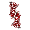
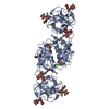
| Structure viewer | Molecule: MolmilJmol/JSmol |
|---|
- Downloads & links
-Download
| PDBx/mmCIF format | 6yh0.cif.gz | 86.7 KB | Display | PDBx/mmCIF format |
|---|---|---|---|---|
| PDB format | pdb6yh0.ent.gz | Display | PDB format | |
| PDBx/mmJSON format | 6yh0.json.gz | Tree view | PDBx/mmJSON format | |
| Others |  Other downloads Other downloads |
-Validation report
| Arichive directory | https://data.pdbj.org/pub/pdb/validation_reports/yh/6yh0ftp://data.pdbj.org/pub/pdb/validation_reports/yh/6yh0 | HTTPS FTP |
|---|
-Related structure data
| Related structure data |  6tslC  6tsmC  6tsnC  6tsoC  6tsqC  6tsrC  3ef2S S: Starting model for refinement C: citing same article ( |
|---|---|
| Similar structure data |


-Links
 PDBj
PDBj

- Assembly
| Deposited unit | 
| ||||||||
|---|---|---|---|---|---|---|---|---|---|
| 1 | 
| ||||||||
| Unit cell |
| ||||||||
| Components on special symmetry positions |
|
-Components
-Protein / Protein/peptide , 2 types, 2 molecules AAAEEE
| #1: Protein | Mass: 32352.559 Da / Num. of mol.: 1 / Mutation: H257A Source method: isolated from a genetically manipulated source Source: (gene. exp.) Marasmius oreades (fairy-ring mushroom)Production host:  Escherichia coli K-12 (bacteria) / References: UniProt: Q8X123 Escherichia coli K-12 (bacteria) / References: UniProt: Q8X123 |
|---|---|
| #2: Protein/peptide | Mass: 764.872 Da / Num. of mol.: 1 / Source method: obtained synthetically / Details: Proteolytic peptide substrate / Source: (synth.) synthetic construct (others) |
-Sugars , 3 types, 3 molecules
| #3: Polysaccharide | alpha-L-fucopyranose-(1-2)-[alpha-D-galactopyranose-(1-3)]beta-D-galactopyranose / Mass: 488.438 Da / Num. of mol.: 1 Source method: isolated from a genetically manipulated source |
|---|---|
| #4: Polysaccharide | alpha-D-galactopyranose-(1-3)-alpha-D-galactopyranose / Mass: 342.297 Da / Num. of mol.: 1 Source method: isolated from a genetically manipulated source |
| #5: Polysaccharide | alpha-L-fucopyranose-(1-2)-[alpha-D-galactopyranose-(1-3)]alpha-D-galactopyranose / Mass: 488.438 Da / Num. of mol.: 1 Source method: isolated from a genetically manipulated source |
-Non-polymers , 4 types, 208 molecules 


| #6: Chemical |  Mass: 40.078 Da / Num. of mol.: 2 / Source method: obtained synthetically / Formula: Ca Mass: 40.078 Da / Num. of mol.: 2 / Source method: obtained synthetically / Formula: Ca#7: Chemical |  Mass: 22.990 Da / Num. of mol.: 2 / Source method: obtained synthetically / Formula: Na Mass: 22.990 Da / Num. of mol.: 2 / Source method: obtained synthetically / Formula: Na#8: Chemical | ChemComp-CL / | Chloride Mass: 35.453 Da / Num. of mol.: 1 / Source method: obtained synthetically / Formula: Cl Mass: 35.453 Da / Num. of mol.: 1 / Source method: obtained synthetically / Formula: Cl#9: Water | ChemComp-HOH / | WaterMass: 18.015 Da / Num. of mol.: 203 / Source method: isolated from a natural source / Formula: H2O |
|---|
-Details
| Has ligand of interest | N |
|---|
-Experimental details
-Experiment
| Experiment | Method: X-RAY DIFFRACTION / Number of used crystals: 1 |
|---|
- Sample preparation
| Crystal | Density Matthews: 3.27 Å3/Da / Density % sol: 62.44 % / Description: rod-like |
|---|---|
| Crystal grow | Temperature: 293.15 K / Method: vapor diffusion, hanging drop / pH: 6.5 Details: 0.1 M Cacodylate pH 6.5, 0.2 M Sodium acetate, 22% PEG 8000, 10 mM CaCl2, 5 mM DTT PH range: 6.5 |
-Data collection
| Diffraction | Mean temperature: 100 K / Ambient temp details: cryostream on a synchrotron beamline / Serial crystal experiment: N |
|---|---|
| Diffraction source | Source: SYNCHROTRON / Site: ESRF  / Beamline: ID29 / Wavelength: 0.972385 Å / Beamline: ID29 / Wavelength: 0.972385 Å |
| Detector | Type: DECTRIS PILATUS 6M / Detector: PIXEL / Date: Jul 20, 2015 |
| Radiation | Monochromator: liquid nitrogen cooled channel-cut silicon monochromator Protocol: SINGLE WAVELENGTH / Monochromatic (M) / Laue (L): M / Scattering type: x-ray |
| Radiation wavelength | Wavelength: 0.972385 Å / Relative weight: 1 |
| Reflection | Resolution: 1.56→105 Å / Num. obs: 61848 / % possible obs: 100 % / Redundancy: 19.6 % / Biso Wilson estimate: 23.9 Å2 / CC1/2: 0.999 / Rmerge(I) obs: 0.048 / Rpim(I) all: 0.037 / Rrim(I) all: 0.166 / Net I/σ(I): 12.3 |
| Reflection shell | Resolution: 1.56→1.59 Å / Redundancy: 15 % / Rmerge(I) obs: 1 / Num. unique obs: 3023 / CC1/2: 0.362 / Rpim(I) all: 1 / Rrim(I) all: 1 / % possible all: 100 |
- Processing
| Software |
| |||||||||||||||||||||||||||||||||||||||||||||||||||||||||||||||||||||||||||||||||||||||||||||||||||||||||||||||||||||||||||||||||||||||||||||||||||||||||||||||||||||
|---|---|---|---|---|---|---|---|---|---|---|---|---|---|---|---|---|---|---|---|---|---|---|---|---|---|---|---|---|---|---|---|---|---|---|---|---|---|---|---|---|---|---|---|---|---|---|---|---|---|---|---|---|---|---|---|---|---|---|---|---|---|---|---|---|---|---|---|---|---|---|---|---|---|---|---|---|---|---|---|---|---|---|---|---|---|---|---|---|---|---|---|---|---|---|---|---|---|---|---|---|---|---|---|---|---|---|---|---|---|---|---|---|---|---|---|---|---|---|---|---|---|---|---|---|---|---|---|---|---|---|---|---|---|---|---|---|---|---|---|---|---|---|---|---|---|---|---|---|---|---|---|---|---|---|---|---|---|---|---|---|---|---|---|---|---|---|
| Refinement | Method to determine structure: MOLECULAR REPLACEMENT Starting model: 3EF2 Resolution: 1.56→51.882 Å / Cor.coef. Fo:Fc: 0.974 / Cor.coef. Fo:Fc free: 0.965 / WRfactor Rfree: 0.177 / WRfactor Rwork: 0.156 / SU B: 1.49 / SU ML: 0.049 / Average fsc free: 0 / Average fsc work: 0 / Cross valid method: FREE R-VALUE / ESU R: 0.065 / ESU R Free: 0.068 Details: Hydrogens have been added in their riding positions
| |||||||||||||||||||||||||||||||||||||||||||||||||||||||||||||||||||||||||||||||||||||||||||||||||||||||||||||||||||||||||||||||||||||||||||||||||||||||||||||||||||||
| Solvent computation | Ion probe radii: 0.8 Å / Shrinkage radii: 0.8 Å / VDW probe radii: 1.2 Å / Solvent model: MASK BULK SOLVENT | |||||||||||||||||||||||||||||||||||||||||||||||||||||||||||||||||||||||||||||||||||||||||||||||||||||||||||||||||||||||||||||||||||||||||||||||||||||||||||||||||||||
| Displacement parameters | Biso mean: 26.788 Å2
| |||||||||||||||||||||||||||||||||||||||||||||||||||||||||||||||||||||||||||||||||||||||||||||||||||||||||||||||||||||||||||||||||||||||||||||||||||||||||||||||||||||
| Refinement step | Cycle: LAST / Resolution: 1.56→51.882 Å
| |||||||||||||||||||||||||||||||||||||||||||||||||||||||||||||||||||||||||||||||||||||||||||||||||||||||||||||||||||||||||||||||||||||||||||||||||||||||||||||||||||||
| Refine LS restraints |
| |||||||||||||||||||||||||||||||||||||||||||||||||||||||||||||||||||||||||||||||||||||||||||||||||||||||||||||||||||||||||||||||||||||||||||||||||||||||||||||||||||||
| LS refinement shell |
|
