 Movie
Movie Controller

[English] 日本語
 Yorodumi
- PDB-6v8i: Composite atomic model of the Staphylococcus aureus phage 80alpha... -
+ Open data

- Basic information
| Entry | Database: PDB / ID: 6v8i | ||||||
|---|---|---|---|---|---|---|---|
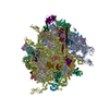
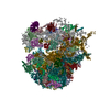
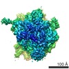
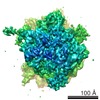
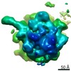
| Title | Composite atomic model of the Staphylococcus aureus phage 80alpha baseplate | ||||||
 Components Components |
| ||||||
 Keywords Keywords |  VIRAL PROTEIN / phage tail / tail tip / tape measure protein VIRAL PROTEIN / phage tail / tail tip / tape measure protein | ||||||
| Function / homology |  Function and homology information Function and homology information | ||||||
| Biological species |  Staphylococcus virus 80alpha Staphylococcus virus 80alpha | ||||||
| Method | ELECTRON MICROSCOPY / single particle reconstruction / cryo EM / Resolution: 3.7 Å | ||||||
 Authors Authors | Kizziah, J.L. / Dokland, T. | ||||||
| Funding support |  United States, 1items United States, 1items
| ||||||
 Citation Citation | Journal: PLoS Pathog / Year: 2020 Title: Structure of the host cell recognition and penetration machinery of a Staphylococcus aureus bacteriophage. Authors: James L Kizziah / Keith A Manning / Altaira D Dearborn / Terje Dokland / Abstract: Staphylococcus aureus is a common cause of infections in humans. The emergence of virulent, antibiotic-resistant strains of S. aureus is a significant public health concern. Most virulence and ...Staphylococcus aureus is a common cause of infections in humans. The emergence of virulent, antibiotic-resistant strains of S. aureus is a significant public health concern. Most virulence and resistance factors in S. aureus are encoded by mobile genetic elements, and transduction by bacteriophages represents the main mechanism for horizontal gene transfer. The baseplate is a specialized structure at the tip of bacteriophage tails that plays key roles in host recognition, cell wall penetration, and DNA ejection. We have used high-resolution cryo-electron microscopy to determine the structure of the S. aureus bacteriophage 80α baseplate at 3.75 Å resolution, allowing atomic models to be built for most of the major tail and baseplate proteins, including two tail fibers, the receptor binding protein, and part of the tape measure protein. Our structure provides a structural basis for understanding host recognition, cell wall penetration and DNA ejection in viruses infecting Gram-positive bacteria. Comparison to other phages demonstrates the modular design of baseplate proteins, and the adaptations to the host that take place during the evolution of staphylococci and other pathogens. | ||||||
| History |
|
- Structure visualization
| Movie |
Movie viewer |
|---|---|
| Structure viewer | Molecule: MolmilJmol/JSmol |
- Downloads & links
-Download
| PDBx/mmCIF format | 6v8i.cif.gz | 3.7 MB | Display | PDBx/mmCIF format |
|---|---|---|---|---|
| PDB format | pdb6v8i.ent.gz | Display | PDB format | |
| PDBx/mmJSON format | 6v8i.json.gz | Tree view | PDBx/mmJSON format | |
| Others |  Other downloads Other downloads |
-Validation report
| Arichive directory | https://data.pdbj.org/pub/pdb/validation_reports/v8/6v8iftp://data.pdbj.org/pub/pdb/validation_reports/v8/6v8i | HTTPS FTP |
|---|
-Related structure data
| Related structure data |  20872MC  20873MC  20874MC  20875MC 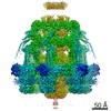 20876MC M: map data used to model this data C: citing same article ( |
|---|---|
| Similar structure data |
-Links
 PDBj
PDBj- Assembly
| Deposited unit | 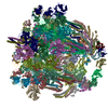
|
|---|---|
| 1 |
|
-Components
-Protein , 7 types, 72 molecules ACADCCCDBCBDAECEBECFBFAFBJBKBLDJDKDLAJAKALFJFKFLCJCKCLEJEKEL...
| #1: Protein | Mass: 37105.004 Da / Num. of mol.: 6 Source method: isolated from a genetically manipulated source Source: (gene. exp.) Staphylococcus virus 80alpha / Strain: ST247 / Production host:   Staphylococcus aureus (bacteria) / References: UniProt: A4ZFC4 Staphylococcus aureus (bacteria) / References: UniProt: A4ZFC4#2: Protein | Mass: 71114.680 Da / Num. of mol.: 3 Source method: isolated from a genetically manipulated source Source: (gene. exp.) Staphylococcus virus 80alpha / Strain: ST247 / Production host: Staphylococcus aureus (bacteria) / References: UniProt: A4ZFC5#3: Protein | Mass: 125891.977 Da / Num. of mol.: 3 Source method: isolated from a genetically manipulated source Source: (gene. exp.) Staphylococcus virus 80alpha / Strain: ST247 / Production host: Staphylococcus aureus (bacteria) / References: UniProt: A4ZFC2#4: Protein | Mass: 66872.477 Da / Num. of mol.: 18 Source method: isolated from a genetically manipulated source Source: (gene. exp.) Staphylococcus virus 80alpha / Strain: ST247 / Production host: Staphylococcus aureus (bacteria) / References: UniProt: A4ZFC8#5: Protein | Mass: 43866.266 Da / Num. of mol.: 12 Source method: isolated from a genetically manipulated source Source: (gene. exp.) Staphylococcus virus 80alpha / Strain: ST247 / Production host: Staphylococcus aureus (bacteria) / References: UniProt: A4ZFD4#6: Protein | Mass: 21551.746 Da / Num. of mol.: 12 Source method: isolated from a genetically manipulated source Source: (gene. exp.) Staphylococcus virus 80alpha / Strain: ST247 / Production host: Staphylococcus aureus (bacteria) / References: UniProt: A4ZFB9#7: Protein | Receptor (biochemistry) / Minor structure proteinMass: 73747.445 Da / Num. of mol.: 18 Source method: isolated from a genetically manipulated source Source: (gene. exp.) Staphylococcus virus 80alpha / Strain: ST247 / Production host: Staphylococcus aureus (bacteria) / References: UniProt: A4ZFC7 |
|---|
-Non-polymers , 1 types, 6 molecules 
| #8: Chemical | ChemComp-FE / Iron Mass: 55.845 Da / Num. of mol.: 6 / Source method: obtained synthetically / Formula: Fe Mass: 55.845 Da / Num. of mol.: 6 / Source method: obtained synthetically / Formula: Fe |
|---|
-Details
| Has ligand of interest | N |
|---|
-Experimental details
-Experiment
| Experiment | Method: ELECTRON MICROSCOPY |
|---|---|
| EM experiment | Aggregation state: PARTICLE / 3D reconstruction method: single particle reconstruction |
- Sample preparation
| Component | Name: Staphylococcus phage 80alpha baseplate / Type: COMPLEX Details: 80alpha baseplate attached to fully formed tails with no capsids Entity ID: #1-#7 / Source: RECOMBINANT | |||||||||||||||||||||||||
|---|---|---|---|---|---|---|---|---|---|---|---|---|---|---|---|---|---|---|---|---|---|---|---|---|---|---|
| Molecular weight | Value: 3.7 MDa / Experimental value: NO | |||||||||||||||||||||||||
| Source (natural) | Organism: Staphylococcus virus 80alpha / Strain: ST247 | |||||||||||||||||||||||||
| Source (recombinant) | Organism: Staphylococcus aureus (bacteria) | |||||||||||||||||||||||||
| Buffer solution | pH: 8 | |||||||||||||||||||||||||
| Buffer component |
| |||||||||||||||||||||||||
| Specimen | Embedding applied: NO / Shadowing applied: NO / Staining applied: NO / Vitrification applied: YES Details: 80alpha tails (with baseplates) purified by centrifugation, polyethylene glycol precipitation, and CsCl density gradation | |||||||||||||||||||||||||
| Specimen support | Grid material: NICKEL / Grid type: Quantifoil R2/1 | |||||||||||||||||||||||||
| Vitrification | Instrument: FEI VITROBOT MARK IV / Cryogen name: ETHANE |
- Electron microscopy imaging
| Experimental equipment |  Model: Titan Krios / Image courtesy: FEI Company |
|---|---|
| Microscopy | Model: FEI TITAN KRIOS |
| Electron gun | Electron source: FIELD EMISSION GUN / Accelerating voltage: 300 kV / Illumination mode: FLOOD BEAM |
| Electron lens | Mode: BRIGHT FIELDBright-field microscopy / Nominal magnification: 29000 X / Nominal defocus max: 3700 nm / Nominal defocus min: 1700 nm |
| Specimen holder | Cryogen: NITROGEN Specimen holder model: GATAN 626 SINGLE TILT LIQUID NITROGEN CRYO TRANSFER HOLDER |
| Image recording | Average exposure time: 1.156 sec. / Electron dose: 104.33 e/Å2 / Film or detector model: DIRECT ELECTRON DE-20 (5k x 3k) / Num. of grids imaged: 10 / Num. of real images: 6483 |
| Image scans | Movie frames/image: 37 / Used frames/image: 1-37 |
- Processing
| EM software |
| |||||||||||||||||||||||||||||||||||||||||||||||||||||||
|---|---|---|---|---|---|---|---|---|---|---|---|---|---|---|---|---|---|---|---|---|---|---|---|---|---|---|---|---|---|---|---|---|---|---|---|---|---|---|---|---|---|---|---|---|---|---|---|---|---|---|---|---|---|---|---|---|
| CTF correction | Type: PHASE FLIPPING ONLY | |||||||||||||||||||||||||||||||||||||||||||||||||||||||
| Particle selection | Num. of particles selected: 515106 | |||||||||||||||||||||||||||||||||||||||||||||||||||||||
| Symmetry | Point symmetry: C6 (6 fold cyclic) | |||||||||||||||||||||||||||||||||||||||||||||||||||||||
| 3D reconstruction | Resolution: 3.7 Å / Resolution method: FSC 0.143 CUT-OFF / Num. of particles: 43601 / Symmetry type: POINT | |||||||||||||||||||||||||||||||||||||||||||||||||||||||
| Atomic model building | Protocol: RIGID BODY FIT / Space: RECIPROCAL / Target criteria: correlation coefficient Details: Initial models derived from I-TASSER predictions based on GenBank sequences of modeled proteins. Rigid body fitting in UCSF Chimera followed by manual extension in Coot. | |||||||||||||||||||||||||||||||||||||||||||||||||||||||
| Atomic model building | PDB-ID: 5EFV Accession code: 5EFV / Source name: PDB / Type: experimental model |

