 Movie
Movie Controller

[English] 日本語
 Yorodumi
- PDB-4v8m: High-resolution cryo-electron microscopy structure of the Trypano... -
+ Open data

- Basic information
| Entry | Database: PDB / ID: 4v8m | |||||||||||||||
|---|---|---|---|---|---|---|---|---|---|---|---|---|---|---|---|---|
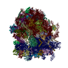
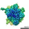
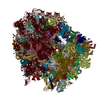
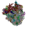
| Title | High-resolution cryo-electron microscopy structure of the Trypanosoma brucei ribosome | |||||||||||||||
 Components Components |
| |||||||||||||||
 Keywords Keywords | RIBOSOME / EUKARYOTIC / KINETOPLASTIDS / EXPANSION SEGMENTS | |||||||||||||||
| Function / homology |  Function and homology information Function and homology informationorganellar small ribosomal subunit / organellar large ribosomal subunit / nuclear lumen / mitochondrial large ribosomal subunit / ciliary transition zone / phosphate ion binding / negative regulation of translational frameshifting / endonucleolytic cleavage to generate mature 3'-end of SSU-rRNA from (SSU-rRNA, 5.8S rRNA, LSU-rRNA) / maturation of LSU-rRNA / protein-RNA complex assembly ...organellar small ribosomal subunit / organellar large ribosomal subunit / nuclear lumen / mitochondrial large ribosomal subunit / ciliary transition zone / phosphate ion binding / negative regulation of translational frameshifting / endonucleolytic cleavage to generate mature 3'-end of SSU-rRNA from (SSU-rRNA, 5.8S rRNA, LSU-rRNA) / maturation of LSU-rRNA / protein-RNA complex assembly / endonucleolytic cleavage in ITS1 to separate SSU-rRNA from 5.8S rRNA and LSU-rRNA from tricistronic rRNA transcript (SSU-rRNA, 5.8S rRNA, LSU-rRNA) / translation regulator activity / rescue of stalled cytosolic ribosome / protein kinase C binding / ribosomal large subunit biogenesis / ribosome assembly / maturation of LSU-rRNA from tricistronic rRNA transcript (SSU-rRNA, 5.8S rRNA, LSU-rRNA) / cytosolic ribosome / maturation of SSU-rRNA / maturation of SSU-rRNA from tricistronic rRNA transcript (SSU-rRNA, 5.8S rRNA, LSU-rRNA) / small-subunit processome / regulation of cell growth / maintenance of translational fidelity / modification-dependent protein catabolic process / protein tag activity / rRNA processing / regulation of cell population proliferation / ribosomal small subunit assembly / ribosome binding / ribosome biogenesis / ribosomal small subunit biogenesis / 5S rRNA binding / ribosomal large subunit assembly / small ribosomal subunit / small ribosomal subunit rRNA binding / cytosolic small ribosomal subunit / large ribosomal subunit rRNA binding / cytosolic large ribosomal subunit / cytoplasmic translation / negative regulation of translation / rRNA binding / protein ubiquitination / structural constituent of ribosome / ribosome / translation / ribonucleoprotein complex / mRNA binding / nucleolus / RNA binding / nucleoplasm / zinc ion binding / metal ion binding / nucleus / cytosol / cytoplasm Similarity search - Function | |||||||||||||||
| Biological species |  | |||||||||||||||
| Method | ELECTRON MICROSCOPY / single particle reconstruction / cryo EM / Resolution: 5.57 Å | |||||||||||||||
 Authors Authors | Hashem, Y. / des Georges, A. / Fu, J. / Buss, S.N. / Jossinet, F. / Jobe, A. / Zhang, Q. / Liao, H.Y. / Grassucci, R.A. / Bajaj, C. ...Hashem, Y. / des Georges, A. / Fu, J. / Buss, S.N. / Jossinet, F. / Jobe, A. / Zhang, Q. / Liao, H.Y. / Grassucci, R.A. / Bajaj, C. / Westhof, E. / Madison-Antenucci, S. / Frank, J. | |||||||||||||||
 Citation Citation | Journal: Nature / Year: 2013 Title: High-resolution cryo-electron microscopy structure of the Trypanosoma brucei ribosome. Authors: Yaser Hashem / Amedee des Georges / Jie Fu / Sarah N Buss / Fabrice Jossinet / Amy Jobe / Qin Zhang / Hstau Y Liao / Robert A Grassucci / Chandrajit Bajaj / Eric Westhof / Susan Madison- ...Authors: Yaser Hashem / Amedee des Georges / Jie Fu / Sarah N Buss / Fabrice Jossinet / Amy Jobe / Qin Zhang / Hstau Y Liao / Robert A Grassucci / Chandrajit Bajaj / Eric Westhof / Susan Madison-Antenucci / Joachim Frank /  Abstract: Ribosomes, the protein factories of living cells, translate genetic information carried by messenger RNAs into proteins, and are thus involved in virtually all aspects of cellular development and ...Ribosomes, the protein factories of living cells, translate genetic information carried by messenger RNAs into proteins, and are thus involved in virtually all aspects of cellular development and maintenance. The few available structures of the eukaryotic ribosome reveal that it is more complex than its prokaryotic counterpart, owing mainly to the presence of eukaryote-specific ribosomal proteins and additional ribosomal RNA insertions, called expansion segments. The structures also differ among species, partly in the size and arrangement of these expansion segments. Such differences are extreme in kinetoplastids, unicellular eukaryotic parasites often infectious to humans. Here we present a high-resolution cryo-electron microscopy structure of the ribosome of Trypanosoma brucei, the parasite that is transmitted by the tsetse fly and that causes African sleeping sickness. The atomic model reveals the unique features of this ribosome, characterized mainly by the presence of unusually large expansion segments and ribosomal-protein extensions leading to the formation of four additional inter-subunit bridges. We also find additional rRNA insertions, including one large rRNA domain that is not found in other eukaryotes. Furthermore, the structure reveals the five cleavage sites of the kinetoplastid large ribosomal subunit (LSU) rRNA chain, which is known to be cleaved uniquely into six pieces, and suggests that the cleavage is important for the maintenance of the T. brucei ribosome in the observed structure. We discuss several possible implications of the large rRNA expansion segments for the translation-regulation process. The structure could serve as a basis for future experiments aimed at understanding the functional importance of these kinetoplastid-specific ribosomal features in protein-translation regulation, an essential step towards finding effective and safe kinetoplastid-specific drugs. | |||||||||||||||
| History |
| |||||||||||||||
| Remark 700 | SHEET DETERMINATION METHOD: DSSP THE SHEETS PRESENTED AS "5B" IN EACH CHAIN ON SHEET RECORDS BELOW ... SHEET DETERMINATION METHOD: DSSP THE SHEETS PRESENTED AS "5B" IN EACH CHAIN ON SHEET RECORDS BELOW IS ACTUALLY AN 5-STRANDED BARREL THIS IS REPRESENTED BY A 6-STRANDED SHEET IN WHICH THE FIRST AND LAST STRANDS ARE IDENTICAL. THE SHEETS PRESENTED AS "EA" IN EACH CHAIN ON SHEET RECORDS BELOW IS ACTUALLY AN 5-STRANDED BARREL THIS IS REPRESENTED BY A 6-STRANDED SHEET IN WHICH THE FIRST AND LAST STRANDS ARE IDENTICAL. THE SHEETS PRESENTED AS "SA" IN EACH CHAIN ON SHEET RECORDS BELOW IS ACTUALLY AN 5-STRANDED BARREL THIS IS REPRESENTED BY A 6-STRANDED SHEET IN WHICH THE FIRST AND LAST STRANDS ARE IDENTICAL. |
- Structure visualization
| Movie |
Movie viewer |
|---|---|
| Structure viewer | Molecule: MolmilJmol/JSmol |
- Downloads & links
-Download
| PDBx/mmCIF format | 4v8m.cif.gz | 5.6 MB | Display | PDBx/mmCIF format |
|---|---|---|---|---|
| PDB format | pdb4v8m.ent.gz | Display | PDB format | |
| PDBx/mmJSON format | 4v8m.json.gz | Tree view | PDBx/mmJSON format | |
| Others |  Other downloads Other downloads |
-Validation report
| Arichive directory | https://data.pdbj.org/pub/pdb/validation_reports/v8/4v8mftp://data.pdbj.org/pub/pdb/validation_reports/v8/4v8m | HTTPS FTP |
|---|
-Related structure data
| Related structure data |  2239MC M: map data used to model this data C: citing same article ( |
|---|---|
| Similar structure data |


-Links
 PDBj
PDBj


- Assembly
| Deposited unit | 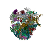
|
|---|---|
| 1 |
|
-Components
+40S RIBOSOMAL PROTEIN ... , 25 types, 25 molecules A0A1A2A3A5A6ACADAFAGAHAIAJAKALAMAPARASATAUAWAXAYAZ
-RIBOSOMAL PROTEIN ... , 9 types, 9 molecules A4A8AOAQAVBQBfBmBn
| #5: Protein | Mass: 23889.201 Da / Num. of mol.: 1 / Source method: isolated from a natural source / Source: (natural) |
|---|---|
| #9: Protein | Mass: 6679.840 Da / Num. of mol.: 1 / Source method: isolated from a natural source / Source: (natural) |
| #22: Protein | Mass: 18891.506 Da / Num. of mol.: 1 / Source method: isolated from a natural source / Source: (natural) |
| #24: Protein | Mass: 13248.478 Da / Num. of mol.: 1 / Source method: isolated from a natural source / Source: (natural) |
| #29: Protein | Mass: 12837.207 Da / Num. of mol.: 1 / Source method: isolated from a natural source / Source: (natural) |
| #50: Protein | Mass: 26461.006 Da / Num. of mol.: 1 / Source method: isolated from a natural source / Source: (natural) |
| #65: Protein | Mass: 48802.996 Da / Num. of mol.: 1 / Source method: isolated from a natural source / Source: (natural) |
| #72: Protein | Mass: 12491.966 Da / Num. of mol.: 1 / Source method: isolated from a natural source / Source: (natural) |
| #73: Protein | Mass: 10128.931 Da / Num. of mol.: 1 / Source method: isolated from a natural source / Source: (natural) |
-Protein , 7 types, 7 molecules A7A9AEBJBPBhBs
| #8: Protein | Mass: 34727.836 Da / Num. of mol.: 1 / Source method: isolated from a natural source / Source: (natural) |
|---|---|
| #10: Protein | Mass: 16936.711 Da / Num. of mol.: 1 / Source method: isolated from a natural source / Source: (natural) |
| #13: Protein | Mass: 20132.459 Da / Num. of mol.: 1 / Source method: isolated from a natural source / Source: (natural) |
| #43: Protein | Mass: 24640.195 Da / Num. of mol.: 1 / Source method: isolated from a natural source / Source: (natural) |
| #49: Protein | Mass: 21742.795 Da / Num. of mol.: 1 / Source method: isolated from a natural source / Source: (natural) |
| #67: Protein | Mass: 21752.922 Da / Num. of mol.: 1 / Source method: isolated from a natural source / Source: (natural) |
| #78: Protein | Mass: 14685.345 Da / Num. of mol.: 1 / Source method: isolated from a natural source / Source: (natural) |
-RNA chain , 10 types, 10 molecules BABBBCBDBEBFBGBHAAAB
| #34: RNA chain | Mass: 595796.438 Da / Num. of mol.: 1 / Source method: isolated from a natural source / Source: (natural) |
|---|---|
| #35: RNA chain | Mass: 471450.062 Da / Num. of mol.: 1 / Source method: isolated from a natural source / Source: (natural) |
| #36: RNA chain | Mass: 54224.051 Da / Num. of mol.: 1 / Source method: isolated from a natural source / Source: (natural) |
| #37: RNA chain | Mass: 38316.672 Da / Num. of mol.: 1 / Source method: isolated from a natural source / Source: (natural) |
| #38: RNA chain | Mass: 67234.633 Da / Num. of mol.: 1 / Source method: isolated from a natural source / Source: (natural) |
| #39: RNA chain | Mass: 23079.566 Da / Num. of mol.: 1 / Source method: isolated from a natural source / Source: (natural) |
| #40: RNA chain | Mass: 58889.910 Da / Num. of mol.: 1 / Source method: isolated from a natural source / Source: (natural) |
| #41: RNA chain | Mass: 43388.613 Da / Num. of mol.: 1 / Source method: isolated from a natural source / Source: (natural) |
| #85: RNA chain | Mass: 724377.062 Da / Num. of mol.: 1 / Source method: isolated from a natural source / Source: (natural) |
| #86: RNA chain | Mass: 23545.971 Da / Num. of mol.: 1 / Source method: isolated from a natural source / Source: (natural) |
+60S RIBOSOMAL PROTEIN ... , 35 types, 35 molecules BIBKBLBMBNBOBRBSBTBUBVBWBXBYBZBaBbBcBdBeBgBiBjBkBlBoBpBqBrBt...
-Details
| Has protein modification | Y |
|---|---|
| Sequence details | CHAINS 8 AND Q ARE MODELLED USING T. CRUZI SEQUENCE UNP ACCESSIONS |
-Experimental details
-Experiment
| Experiment | Method: ELECTRON MICROSCOPY |
|---|---|
| EM experiment | Aggregation state: PARTICLE / 3D reconstruction method: single particle reconstruction |
- Sample preparation
| Component | Name: TRYPANOSOMA BRUCEI 80S RIBOSOME / Type: RIBOSOME / Details: MICROGRAPHS SELECTED VISUALLY AFTER SCANNING |
|---|---|
| Buffer solution | Name: 20 MM HEPES, 10MM MGCL2, 500 MM KCL AND 5 MM BETA-ME / pH: 7.2 Details: 20 MM HEPES, 10MM MGCL2, 500 MM KCL AND 5 MM BETA-ME |
| Specimen | Embedding applied: NO / Shadowing applied: NO / Staining applied: NO / Vitrification applied: YES |
| Specimen support | Details: CARBON |
| Vitrification | Instrument: FEI VITROBOT MARK IV / Cryogen name: ETHANE / Details: LIQUID ETHAN |
- Electron microscopy imaging
| Experimental equipment |  Model: Tecnai F30 / Image courtesy: FEI Company |
|---|---|
| Microscopy | Model: FEI TECNAI F30 / Date: Jan 1, 2011 Details: MICROGRAPHS SCANNED WITH NIKON SUPER COOLSCAN 9000ED |
| Electron gun | Electron source:  FIELD EMISSION GUN / Accelerating voltage: 300 kV / Illumination mode: FLOOD BEAM FIELD EMISSION GUN / Accelerating voltage: 300 kV / Illumination mode: FLOOD BEAM |
| Electron lens | Mode: BRIGHT FIELD / Nominal magnification: 59000 X / Nominal defocus max: 4000 nm / Nominal defocus min: 1500 nm / Cs: 2 mm |
| Specimen holder | Temperature: 93.15 K |
| Image recording | Electron dose: 25 e/Å2 / Film or detector model: KODAK SO-163 FILM |
| Image scans | Num. digital images: 1102 |
| Radiation wavelength | Relative weight: 1 |
- Processing
| EM software |
| |||||||||||||||||||||||||||||||||||||||||||||||||||||||||||||||||||||||||||||||||||||||||||||||||||||||||||||||||||||||||
|---|---|---|---|---|---|---|---|---|---|---|---|---|---|---|---|---|---|---|---|---|---|---|---|---|---|---|---|---|---|---|---|---|---|---|---|---|---|---|---|---|---|---|---|---|---|---|---|---|---|---|---|---|---|---|---|---|---|---|---|---|---|---|---|---|---|---|---|---|---|---|---|---|---|---|---|---|---|---|---|---|---|---|---|---|---|---|---|---|---|---|---|---|---|---|---|---|---|---|---|---|---|---|---|---|---|---|---|---|---|---|---|---|---|---|---|---|---|---|---|---|---|---|
| CTF correction | Details: PHASE-FLIPPING | |||||||||||||||||||||||||||||||||||||||||||||||||||||||||||||||||||||||||||||||||||||||||||||||||||||||||||||||||||||||||
| Symmetry | Point symmetry: C1 (asymmetric) | |||||||||||||||||||||||||||||||||||||||||||||||||||||||||||||||||||||||||||||||||||||||||||||||||||||||||||||||||||||||||
| 3D reconstruction | Method: REFERENCE BASED RECONSTRUCTION METHOD / Resolution: 5.57 Å / Num. of particles: 164000 / Nominal pixel size: 1.09 Å Details: SUBMISSION BASED ON EXPERIMENTAL DATA FROM EMDB EMD-2239 (DEPOSITION ID: 11259). THE STRUCTURE OF THE 80S FROM YEAST (3U5B AND OTHERS) AS WELL AS THE STRUCTURE OF THE 60S FROM TETRAHYMENA ...Details: SUBMISSION BASED ON EXPERIMENTAL DATA FROM EMDB EMD-2239 (DEPOSITION ID: 11259). THE STRUCTURE OF THE 80S FROM YEAST (3U5B AND OTHERS) AS WELL AS THE STRUCTURE OF THE 60S FROM TETRAHYMENA THERMOPHILA (4A17 AND OTHERS) WERE USED AS STARTING MODEL FOR THE 60S SUBUNIT MODEL. THE 40S FROM TETRAHYMENA THERMOPHILA (2XZM AND 2XZN) AS WELL AS THE 80S FROM YEAST WERE USED AS STARTING MODEL FOR THE 40S SUBUNIT MODEL. THE 80S MODEL OF TRITICUM AESTIVUM (3IZR AND OTHERS) WAS USED TO FIT MISSING PROTEINS FORM THE TWO X-RAY STRUCTURES. Symmetry type: POINT | |||||||||||||||||||||||||||||||||||||||||||||||||||||||||||||||||||||||||||||||||||||||||||||||||||||||||||||||||||||||||
| Atomic model building | Protocol: RIGID BODY FIT / Space: REAL / Details: REFINEMENT PROTOCOL--RIGID BODY | |||||||||||||||||||||||||||||||||||||||||||||||||||||||||||||||||||||||||||||||||||||||||||||||||||||||||||||||||||||||||
| Atomic model building |
|
