 Movie
Movie Controller


+ Open data

- Basic information
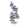
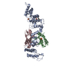
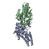
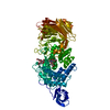
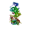
| Entry | Database: PDB / ID: 6sc6 | ||||||
|---|---|---|---|---|---|---|---|
| Title | dAb3/HOIP-RBR apo structure | ||||||
 Components Components |
| ||||||
 Keywords Keywords | LIGASE / Human Single Domain Antibody / HOIP / RBR | ||||||
| Function / homology |  Function and homology information Function and homology informationprotein linear polyubiquitination / LUBAC complex / linear polyubiquitin binding / RBR-type E3 ubiquitin transferase / CD40 signaling pathway / positive regulation of xenophagy / CD40 receptor complex / negative regulation of necroptotic process / TNFR1-induced proapoptotic signaling / K48-linked polyubiquitin modification-dependent protein binding ...protein linear polyubiquitination / LUBAC complex / linear polyubiquitin binding / RBR-type E3 ubiquitin transferase / CD40 signaling pathway / positive regulation of xenophagy / CD40 receptor complex / negative regulation of necroptotic process / TNFR1-induced proapoptotic signaling / K48-linked polyubiquitin modification-dependent protein binding / : / K63-linked polyubiquitin modification-dependent protein binding / ubiquitin binding / TNFR1-induced NF-kappa-B signaling pathway / Regulation of TNFR1 signaling / : / cytoplasmic side of plasma membrane / protein polyubiquitination / ubiquitin-protein transferase activity / ubiquitin protein ligase activity / T cell receptor signaling pathway / positive regulation of canonical NF-kappaB signal transduction / defense response to bacterium / ubiquitin protein ligase binding / zinc ion binding / identical protein binding / cytosol Similarity search - Function | ||||||
| Biological species |  Homo sapiens (human) Homo sapiens (human)synthetic construct (others) | ||||||
| Method |  X-RAY DIFFRACTION / SYNCHROTRON / MOLECULAR REPLACEMENT / Resolution: 2.25 Å X-RAY DIFFRACTION / SYNCHROTRON / MOLECULAR REPLACEMENT / Resolution: 2.25 Å | ||||||
 Authors Authors | Tsai, Y.-C.I. / House, D. / Rittinger, K. | ||||||
| Funding support |  United Kingdom, 1items United Kingdom, 1items
| ||||||
 Citation Citation | Journal: Cell Chem Biol / Year: 2020 Title: Single-Domain Antibodies as Crystallization Chaperones to Enable Structure-Based Inhibitor Development for RBR E3 Ubiquitin Ligases. Authors: Tsai, Y.I. / Johansson, H. / Dixon, D. / Martin, S. / Chung, C.W. / Clarkson, J. / House, D. / Rittinger, K. | ||||||
| History |
|
- Structure visualization
| Structure viewer | Molecule: MolmilJmol/JSmol |
|---|
- Downloads & links
-Download
| PDBx/mmCIF format | 6sc6.cif.gz | 260.6 KB | Display | PDBx/mmCIF format |
|---|---|---|---|---|
| PDB format | pdb6sc6.ent.gz | 208.8 KB | Display | PDB format |
| PDBx/mmJSON format | 6sc6.json.gz | Tree view | PDBx/mmJSON format | |
| Others |  Other downloads Other downloads |
-Validation report
| Arichive directory | https://data.pdbj.org/pub/pdb/validation_reports/sc/6sc6ftp://data.pdbj.org/pub/pdb/validation_reports/sc/6sc6 | HTTPS FTP |
|---|
-Related structure data
| Related structure data |  6sc5C  6sc7C 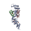 6sc8C  6sc9C 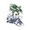 6t2jC 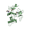 4ljqS  6sca S: Starting model for refinement C: citing same article ( |
|---|---|
| Similar structure data |


-Links
 PDBj
PDBj


- Assembly
| Deposited unit | 
| |||||||||||||||||||||||||||||||||||||||||||||||||||||||||||||||||||||||||||||||||||||||||||||
|---|---|---|---|---|---|---|---|---|---|---|---|---|---|---|---|---|---|---|---|---|---|---|---|---|---|---|---|---|---|---|---|---|---|---|---|---|---|---|---|---|---|---|---|---|---|---|---|---|---|---|---|---|---|---|---|---|---|---|---|---|---|---|---|---|---|---|---|---|---|---|---|---|---|---|---|---|---|---|---|---|---|---|---|---|---|---|---|---|---|---|---|---|---|---|
| 1 |
| |||||||||||||||||||||||||||||||||||||||||||||||||||||||||||||||||||||||||||||||||||||||||||||
| Unit cell |
| |||||||||||||||||||||||||||||||||||||||||||||||||||||||||||||||||||||||||||||||||||||||||||||
| Noncrystallographic symmetry (NCS) | NCS domain:
NCS domain segments: Ens-ID: 1
|