 Movie
Movie Controller

[English] 日本語
 Yorodumi
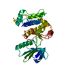
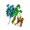
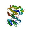
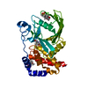
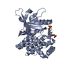
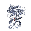
- PDB-6qfl: Structure of the mitogen activated kinase kinase 7 active conformation -
+ Open data

- Basic information
| Entry | Database: PDB / ID: 6qfl | ||||||
|---|---|---|---|---|---|---|---|
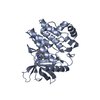
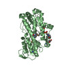
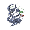
| Title | Structure of the mitogen activated kinase kinase 7 active conformation | ||||||
 Components Components | Dual specificity mitogen-activated protein kinase kinase 7 | ||||||
 Keywords Keywords |  TRANSFERASE / Protein Kinase / Kinase / MKK7 TRANSFERASE / Protein Kinase / Kinase / MKK7 | ||||||
| Function / homology |  Function and homology information Function and homology informationJUN kinase kinase activity / regulation of motor neuron apoptotic process / mitogen-activated protein kinase kinase / response to osmotic stress / Fc-epsilon receptor signaling pathway / positive regulation of telomere capping / MAP kinase kinase activity / Uptake and function of anthrax toxins / MAP kinase activity / cellular response to interleukin-1 ...JUN kinase kinase activity / regulation of motor neuron apoptotic process / mitogen-activated protein kinase kinase / response to osmotic stress / Fc-epsilon receptor signaling pathway / positive regulation of telomere capping / MAP kinase kinase activity / Uptake and function of anthrax toxins / MAP kinase activity / cellular response to interleukin-1 / response to tumor necrosis factor / stress-activated MAPK cascade / response to UV / positive regulation of JUN kinase activity / JNK cascade / positive regulation of telomerase activity / positive regulation of telomere maintenance via telomerase / JNK (c-Jun kinases) phosphorylation and activation mediated by activated human TAK1 / molecular function activator activity / FCERI mediated MAPK activation / positive regulation of JNK cascade / response to wounding / cellular senescence / response to heat / protein phosphatase binding / protein tyrosine kinase activity / Oxidative Stress Induced Senescence / cellular response to lipopolysaccharide / positive regulation of ERK1 and ERK2 cascade / phosphorylation / protein serine kinase activity / apoptotic process / protein kinase binding / positive regulation of DNA-templated transcription / enzyme binding / magnesium ion binding / signal transduction / ATP binding / nucleus / cytosol / cytoplasmSimilarity search - Function | ||||||
| Biological species |  Homo sapiens (human) Homo sapiens (human) | ||||||
| Method | X-RAY DIFFRACTION / SYNCHROTRON / MOLECULAR REPLACEMENT / Resolution: 2.2 Å | ||||||
 Authors Authors | Wolle, P. / Mueller, M.P. / Rauh, D. | ||||||
 Citation Citation | Journal: J.Med.Chem. / Year: 2019 Title: Characterization of Covalent Pyrazolopyrimidine-MKK7 Complexes and a Report on a Unique DFG-in/Leu-in Conformation of Mitogen-Activated Protein Kinase Kinase 7 (MKK7). Authors: Wolle, P. / Engel, J. / Smith, S. / Goebel, L. / Hennes, E. / Lategahn, J. / Rauh, D. | ||||||
| History |
|
- Structure visualization
| Structure viewer | Molecule: MolmilJmol/JSmol |
|---|
- Downloads & links
-Download
| PDBx/mmCIF format | 6qfl.cif.gz | 71.9 KB | Display | PDBx/mmCIF format |
|---|---|---|---|---|
| PDB format | pdb6qfl.ent.gz | 50.7 KB | Display | PDB format |
| PDBx/mmJSON format | 6qfl.json.gz | Tree view | PDBx/mmJSON format | |
| Others |  Other downloads Other downloads |
-Validation report
| Arichive directory | https://data.pdbj.org/pub/pdb/validation_reports/qf/6qflftp://data.pdbj.org/pub/pdb/validation_reports/qf/6qfl | HTTPS FTP |
|---|
-Related structure data
| Related structure data | 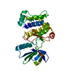 6qfrC 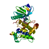 6qftC  6qg4C 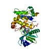 6qg7C 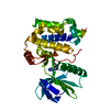 6qhoC 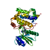 6qhrC 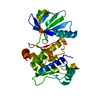 2dylS S: Starting model for refinement C: citing same article ( |
|---|---|
| Similar structure data |

-Links
 PDBj
PDBj


- Assembly
| Deposited unit | 
| ||||||||
|---|---|---|---|---|---|---|---|---|---|
| 1 |
| ||||||||
| Unit cell |
|
-Components
| #1: Protein | Mass: 36146.859 Da / Num. of mol.: 1 Source method: isolated from a genetically manipulated source Source: (gene. exp.) Homo sapiens (human) / Gene: MAP2K7, JNKK2, MEK7, MKK7, PRKMK7, SKK4 / Production host:  Escherichia coli (E. coli) / Strain (production host): BL21 DE3 Escherichia coli (E. coli) / Strain (production host): BL21 DE3References: UniProt: O14733, mitogen-activated protein kinase kinase |
|---|---|
| #2: Water | ChemComp-HOH / Water Mass: 18.015 Da / Num. of mol.: 72 / Source method: isolated from a natural source / Formula: H2O Mass: 18.015 Da / Num. of mol.: 72 / Source method: isolated from a natural source / Formula: H2O |
-Experimental details
-Experiment
| Experiment | Method: X-RAY DIFFRACTION / Number of used crystals: 1 |
|---|
- Sample preparation
| Crystal | Density Matthews: 2.78 Å3/Da / Density % sol: 55.73 % |
|---|---|
| Crystal grow | Temperature: 277 K / Method: vapor diffusion, hanging drop / Details: 0.2 M Sodiumcitrate 20 % PEG3350 |
-Data collection
| Diffraction | Mean temperature: 100 K / Serial crystal experiment: N |
|---|---|
| Diffraction source | Source: SYNCHROTRON / Site: SLS  / Beamline: X10SA / Wavelength: 0.9786 Å / Beamline: X10SA / Wavelength: 0.9786 Å |
| Detector | Type: DECTRIS PILATUS 6M / Detector: PIXEL / Date: Aug 21, 2015 |
| Radiation | Protocol: SINGLE WAVELENGTH / Monochromatic (M) / Laue (L): M / Scattering type: x-ray |
| Radiation wavelength | Wavelength: 0.9786 Å / Relative weight: 1 |
| Reflection | Resolution: 2.2→46.058 Å / Num. obs: 19157 / % possible obs: 92.8 % / Redundancy: 5.8 % / CC1/2: 0.997 / Rrim(I) all: 0.075 / Rsym value: 0.067 / Net I/σ(I): 17.11 |
| Reflection shell | Resolution: 2.2→2.4 Å / Redundancy: 5.93 % / Mean I/σ(I) obs: 3.54 / Num. unique obs: 3534 / CC1/2: 0.994 / Rrim(I) all: 0.447 / Rsym value: 0.402 / % possible all: 81.8 |
- Processing
| Software |
| |||||||||||||||||||||||||||||||||||||||||||||||||||||||||||||||||||||||||||||
|---|---|---|---|---|---|---|---|---|---|---|---|---|---|---|---|---|---|---|---|---|---|---|---|---|---|---|---|---|---|---|---|---|---|---|---|---|---|---|---|---|---|---|---|---|---|---|---|---|---|---|---|---|---|---|---|---|---|---|---|---|---|---|---|---|---|---|---|---|---|---|---|---|---|---|---|---|---|---|
| Refinement | Method to determine structure: MOLECULAR REPLACEMENT Starting model: 2DYL Resolution: 2.2→46.058 Å / SU ML: 0.29 / Cross valid method: FREE R-VALUE / σ(F): 1.36 / Phase error: 30.55
| |||||||||||||||||||||||||||||||||||||||||||||||||||||||||||||||||||||||||||||
| Solvent computation | Shrinkage radii: 0.9 Å / VDW probe radii: 1.11 Å | |||||||||||||||||||||||||||||||||||||||||||||||||||||||||||||||||||||||||||||
| Refinement step | Cycle: LAST / Resolution: 2.2→46.058 Å
| |||||||||||||||||||||||||||||||||||||||||||||||||||||||||||||||||||||||||||||
| Refine LS restraints |
| |||||||||||||||||||||||||||||||||||||||||||||||||||||||||||||||||||||||||||||
| LS refinement shell |
|
