 Movie
Movie Controller

[English] 日本語
 Yorodumi
- PDB-6q58: Copper loading to a cytosolic copper storage protein from Strepto... -
+ Open data

- Basic information
| Entry | Database: PDB / ID: 6q58 | ||||||
|---|---|---|---|---|---|---|---|
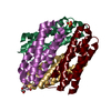
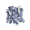
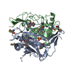
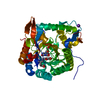
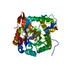
| Title | Copper loading to a cytosolic copper storage protein from Streptomyces lividans (five coppers) | ||||||
 Components Components | Cytosolic copper storage protein | ||||||
 Keywords Keywords | METAL BINDING PROTEIN / Copper / cytosolic / storage protein / Streptomyces livdians | ||||||
| Function / homology | Protein of unknown function DUF326 / Domain of Unknown Function (DUF326) / Uncharacterized cysteine-rich protein YhjQ-like / metal ion binding / COPPER (I) ION / Four-helix bundle copper-binding protein Function and homology information Function and homology information | ||||||
| Biological species |  Streptomyces coelicolor 1326 (bacteria) Streptomyces coelicolor 1326 (bacteria) | ||||||
| Method |  X-RAY DIFFRACTION / SYNCHROTRON / MOLECULAR REPLACEMENT / Resolution: 1.5 Å X-RAY DIFFRACTION / SYNCHROTRON / MOLECULAR REPLACEMENT / Resolution: 1.5 Å | ||||||
 Authors Authors | Straw, M.L. / Hough, M.A. / Worrall, J.A.R. | ||||||
 Citation Citation | Journal: Chemistry / Year: 2019 Title: A Histidine Residue and a Tetranuclear Cuprous-thiolate Cluster Dominate the Copper Loading Landscape of a Copper Storage Protein from Streptomyces lividans. Authors: Straw, M.L. / Hough, M.A. / Wilson, M.T. / Worrall, J.A.R. | ||||||
| History |
|
- Structure visualization
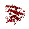
| Structure viewer | Molecule: MolmilJmol/JSmol |
|---|
- Downloads & links
-Download
| PDBx/mmCIF format | 6q58.cif.gz | 108.6 KB | Display | PDBx/mmCIF format |
|---|---|---|---|---|
| PDB format | pdb6q58.ent.gz | 82.5 KB | Display | PDB format |
| PDBx/mmJSON format | 6q58.json.gz | Tree view | PDBx/mmJSON format | |
| Others |  Other downloads Other downloads |
-Validation report
| Summary document | 6q58_validation.pdf.gz | 444.1 KB | Display | wwPDB validaton report |
|---|---|---|---|---|
| Full document | 6q58_full_validation.pdf.gz | 447.2 KB | Display | |
| Data in XML | 6q58_validation.xml.gz | 23.1 KB | Display | |
| Data in CIF | 6q58_validation.cif.gz | 34 KB | Display | |
| Arichive directory | https://data.pdbj.org/pub/pdb/validation_reports/q5/6q58ftp://data.pdbj.org/pub/pdb/validation_reports/q5/6q58 | HTTPS FTP |
-Related structure data
| Related structure data |  6q6bC 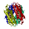 6qvhC 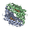 6qybC  6r01C  6ei0S S: Starting model for refinement C: citing same article ( |
|---|---|
| Similar structure data |


-Links
 PDBj
PDBj- Assembly
| Deposited unit | 
| ||||||||||||||||||||||||||||||||||||||||||||||||||||||||||||||||||||||||||||||||||||||||||||||||||||||||||||||||||||||||||||||||||||||||||||||||||||||||||||||||||||||||||||||||
|---|---|---|---|---|---|---|---|---|---|---|---|---|---|---|---|---|---|---|---|---|---|---|---|---|---|---|---|---|---|---|---|---|---|---|---|---|---|---|---|---|---|---|---|---|---|---|---|---|---|---|---|---|---|---|---|---|---|---|---|---|---|---|---|---|---|---|---|---|---|---|---|---|---|---|---|---|---|---|---|---|---|---|---|---|---|---|---|---|---|---|---|---|---|---|---|---|---|---|---|---|---|---|---|---|---|---|---|---|---|---|---|---|---|---|---|---|---|---|---|---|---|---|---|---|---|---|---|---|---|---|---|---|---|---|---|---|---|---|---|---|---|---|---|---|---|---|---|---|---|---|---|---|---|---|---|---|---|---|---|---|---|---|---|---|---|---|---|---|---|---|---|---|---|---|---|---|---|
| 1 | 
| ||||||||||||||||||||||||||||||||||||||||||||||||||||||||||||||||||||||||||||||||||||||||||||||||||||||||||||||||||||||||||||||||||||||||||||||||||||||||||||||||||||||||||||||||
| Unit cell |
| ||||||||||||||||||||||||||||||||||||||||||||||||||||||||||||||||||||||||||||||||||||||||||||||||||||||||||||||||||||||||||||||||||||||||||||||||||||||||||||||||||||||||||||||||
| Noncrystallographic symmetry (NCS) | NCS domain:
NCS domain segments: Component-ID: _ / Refine code: _
NCS ensembles :
|
-Components
| #1: Protein | Mass: 12535.339 Da / Num. of mol.: 4 Source method: isolated from a genetically manipulated source Source: (gene. exp.) Streptomyces coelicolor 1326 (bacteria)Gene: SCO3281 / Production host: #2: Chemical | ChemComp-CU1 /   Mass: 63.546 Da / Num. of mol.: 15 / Source method: obtained synthetically / Formula: Cu Mass: 63.546 Da / Num. of mol.: 15 / Source method: obtained synthetically / Formula: Cu#3: Water | ChemComp-HOH / |  Mass: 18.015 Da / Num. of mol.: 423 / Source method: isolated from a natural source / Formula: H2O Mass: 18.015 Da / Num. of mol.: 423 / Source method: isolated from a natural source / Formula: H2O |
|---|
-Experimental details
-Experiment
| Experiment | Method: X-RAY DIFFRACTION / Number of used crystals: 1 |
|---|
- Sample preparation
| Crystal | Density Matthews: 2.65 Å3/Da / Density % sol: 53.52 % |
|---|---|
| Crystal grow | Temperature: 293 K / Method: vapor diffusion, hanging drop / Details: 1.5 M Ammonium Sulfate 0.1M MES pH 6.0 |
-Data collection
| Diffraction | Mean temperature: 100 K / Serial crystal experiment: N | ||||||||||||||||||||||||
|---|---|---|---|---|---|---|---|---|---|---|---|---|---|---|---|---|---|---|---|---|---|---|---|---|---|
| Diffraction source | Source: SYNCHROTRON / Site: SLS  / Beamline: X10SA / Wavelength: 1.33 Å / Beamline: X10SA / Wavelength: 1.33 Å | ||||||||||||||||||||||||
| Detector | Type: DECTRIS PILATUS 6M-F / Detector: PIXEL / Date: Nov 11, 2016 | ||||||||||||||||||||||||
| Radiation | Protocol: SINGLE WAVELENGTH / Monochromatic (M) / Laue (L): M / Scattering type: x-ray | ||||||||||||||||||||||||
| Radiation wavelength | Wavelength: 1.33 Å / Relative weight: 1 | ||||||||||||||||||||||||
| Reflection | Resolution: 1.5→212.34 Å / Num. obs: 85587 / % possible obs: 98 % / Redundancy: 8 % / CC1/2: 0.999 / Rmerge(I) obs: 0.057 / Rpim(I) all: 0.02 / Rrim(I) all: 0.06 / Net I/σ(I): 19.5 | ||||||||||||||||||||||||
| Reflection shell | Diffraction-ID: 1
|
- Processing
| Software |
| |||||||||||||||||||||||||||||||||||||||||||||||||||||||||||||||||
|---|---|---|---|---|---|---|---|---|---|---|---|---|---|---|---|---|---|---|---|---|---|---|---|---|---|---|---|---|---|---|---|---|---|---|---|---|---|---|---|---|---|---|---|---|---|---|---|---|---|---|---|---|---|---|---|---|---|---|---|---|---|---|---|---|---|---|
| Refinement | Method to determine structure: MOLECULAR REPLACEMENT Starting model: 6EI0 Resolution: 1.5→212.34 Å / Cor.coef. Fo:Fc: 0.963 / Cor.coef. Fo:Fc free: 0.951 / SU B: 1.463 / SU ML: 0.053 / SU R Cruickshank DPI: 0.0724 / Cross valid method: THROUGHOUT / σ(F): 0 / ESU R: 0.072 / ESU R Free: 0.072 Details: HYDROGENS HAVE BEEN ADDED IN THE RIDING POSITIONS U VALUES : REFINED INDIVIDUALLY
| |||||||||||||||||||||||||||||||||||||||||||||||||||||||||||||||||
| Solvent computation | Ion probe radii: 0.8 Å / Shrinkage radii: 0.8 Å / VDW probe radii: 1.2 Å | |||||||||||||||||||||||||||||||||||||||||||||||||||||||||||||||||
| Displacement parameters | Biso max: 97.19 Å2 / Biso mean: 27.975 Å2 / Biso min: 13.39 Å2
| |||||||||||||||||||||||||||||||||||||||||||||||||||||||||||||||||
| Refinement step | Cycle: final / Resolution: 1.5→212.34 Å
| |||||||||||||||||||||||||||||||||||||||||||||||||||||||||||||||||
| Refine LS restraints |
| |||||||||||||||||||||||||||||||||||||||||||||||||||||||||||||||||
| Refine LS restraints NCS | Refine-ID: X-RAY DIFFRACTION / Type: interatomic distance / Weight position: 0.05
| |||||||||||||||||||||||||||||||||||||||||||||||||||||||||||||||||
| LS refinement shell | Resolution: 1.5→1.539 Å / Rfactor Rfree error: 0 / Total num. of bins used: 20
|
