 Movie
Movie Controller

[English] 日本語
 Yorodumi
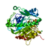
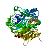
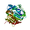
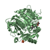
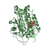
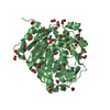
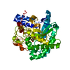
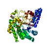
- PDB-5lz2: Fragment-based inhibitors of Lipoprotein associated Phospholipase A2 -
+ Open data

- Basic information
| Entry | Database: PDB / ID: 5lz2 | ||||||
|---|---|---|---|---|---|---|---|
| Title | Fragment-based inhibitors of Lipoprotein associated Phospholipase A2 | ||||||
 Components Components | Platelet-activating factor acetylhydrolase | ||||||
 Keywords Keywords | HYDROLASE / Lp-PLA2 phospholipase / lipid metabolism / inhibitors / Lp-PLA2#4 | ||||||
| Function / homology |  Function and homology information Function and homology informationplasma lipoprotein particle oxidation / platelet activating factor catabolic process / : / 1-alkyl-2-acetylglycerophosphocholine esterase / 1-alkyl-2-acetylglycerophosphocholine esterase activity / platelet activating factor metabolic process / lipid oxidation / low-density lipoprotein particle / high-density lipoprotein particle / low-density lipoprotein particle remodeling ...plasma lipoprotein particle oxidation / platelet activating factor catabolic process / : / 1-alkyl-2-acetylglycerophosphocholine esterase / 1-alkyl-2-acetylglycerophosphocholine esterase activity / platelet activating factor metabolic process / lipid oxidation / low-density lipoprotein particle / high-density lipoprotein particle / low-density lipoprotein particle remodeling / phosphatidylcholine catabolic process / positive regulation of monocyte chemotaxis / peptide hormone processing / hydrolase activity, acting on ester bonds / Synthesis, secretion, and deacylation of Ghrelin / phospholipid binding / positive regulation of inflammatory response / extracellular region Similarity search - Function | ||||||
| Biological species |  Homo sapiens (human) Homo sapiens (human) | ||||||
| Method |  X-RAY DIFFRACTION / FOURIER SYNTHESIS / Resolution: 2.1 Å X-RAY DIFFRACTION / FOURIER SYNTHESIS / Resolution: 2.1 Å | ||||||
 Authors Authors | Woolford, A. / Day, P. | ||||||
 Citation Citation | Journal: J. Med. Chem. / Year: 2016 Title: Fragment-Based Approach to the Development of an Orally Bioavailable Lactam Inhibitor of Lipoprotein-Associated Phospholipase A2 (Lp-PLA2). Authors: Woolford, A.J. / Day, P.J. / Beneton, V. / Berdini, V. / Coyle, J.E. / Dudit, Y. / Grondin, P. / Huet, P. / Lee, L.Y. / Manas, E.S. / McMenamin, R.L. / Murray, C.W. / Page, L.W. / Patel, V.K. ...Authors: Woolford, A.J. / Day, P.J. / Beneton, V. / Berdini, V. / Coyle, J.E. / Dudit, Y. / Grondin, P. / Huet, P. / Lee, L.Y. / Manas, E.S. / McMenamin, R.L. / Murray, C.W. / Page, L.W. / Patel, V.K. / Potvain, F. / Rich, S.J. / Sang, Y. / Somers, D.O. / Trottet, L. / Wan, Z. / Zhang, X. | ||||||
| History |
|
- Structure visualization
| Structure viewer | Molecule: MolmilJmol/JSmol |
|---|
- Downloads & links
-Download
| PDBx/mmCIF format | 5lz2.cif.gz | 174.8 KB | Display | PDBx/mmCIF format |
|---|---|---|---|---|
| PDB format | pdb5lz2.ent.gz | 136.3 KB | Display | PDB format |
| PDBx/mmJSON format | 5lz2.json.gz | Tree view | PDBx/mmJSON format | |
| Others |  Other downloads Other downloads |
-Validation report
| Arichive directory | https://data.pdbj.org/pub/pdb/validation_reports/lz/5lz2ftp://data.pdbj.org/pub/pdb/validation_reports/lz/5lz2 | HTTPS FTP |
|---|
-Related structure data
| Related structure data | 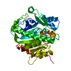 5lyyC 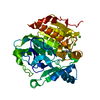 5lz4C 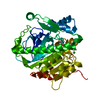 5lz5C 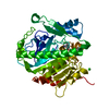 5lz7C 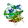 5lz8C  5lz9C C: citing same article ( |
|---|---|
| Similar structure data |


-Links
 PDBj
PDBj


- Assembly
| Deposited unit | 
| ||||||||||||||||||
|---|---|---|---|---|---|---|---|---|---|---|---|---|---|---|---|---|---|---|---|
| 1 |
| ||||||||||||||||||
| Unit cell |
| ||||||||||||||||||
| Components on special symmetry positions |
|
-Components
| #1: Protein | Mass: 44203.129 Da / Num. of mol.: 1 Source method: isolated from a genetically manipulated source Source: (gene. exp.) Homo sapiens (human) / Gene: PLA2G7, PAFAH / Production host:  References: UniProt: Q13093, 1-alkyl-2-acetylglycerophosphocholine esterase |
|---|---|
| #2: Chemical | ChemComp-NA /   Mass: 22.990 Da / Num. of mol.: 1 / Source method: obtained synthetically / Formula: Na Mass: 22.990 Da / Num. of mol.: 1 / Source method: obtained synthetically / Formula: Na |
| #3: Chemical | ChemComp-CL /   Mass: 35.453 Da / Num. of mol.: 1 / Source method: obtained synthetically / Formula: Cl Mass: 35.453 Da / Num. of mol.: 1 / Source method: obtained synthetically / Formula: Cl |
| #4: Chemical | ChemComp-7BX / 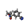  Mass: 222.264 Da / Num. of mol.: 1 / Source method: obtained synthetically / Formula: C10H10N2O2S Mass: 222.264 Da / Num. of mol.: 1 / Source method: obtained synthetically / Formula: C10H10N2O2S |
| #5: Water | ChemComp-HOH /  Mass: 18.015 Da / Num. of mol.: 349 / Source method: isolated from a natural source / Formula: H2O Mass: 18.015 Da / Num. of mol.: 349 / Source method: isolated from a natural source / Formula: H2O |
-Experimental details
-Experiment
| Experiment | Method: X-RAY DIFFRACTION / Number of used crystals: 1 |
|---|
- Sample preparation
| Crystal | Density Matthews: 2.44 Å3/Da / Density % sol: 49.63 % |
|---|---|
| Crystal grow | Temperature: 293 K / Method: vapor diffusion, sitting drop / pH: 7.4 Details: 28.0%w/v PEG 3350, 0.1M HEPES/NaOHpH=7.4, 1.3M NaCl, |
-Data collection
| Diffraction | Mean temperature: 100 K |
|---|---|
| Diffraction source | Source: ROTATING ANODE / Type: RIGAKU FR-E SUPERBRIGHT / Wavelength: 1.5418 Å |
| Detector | Type: RIGAKU RAXIS IV / Detector: IMAGE PLATE / Date: Sep 17, 2010 / Details: mirrors |
| Radiation | Protocol: SINGLE WAVELENGTH / Monochromatic (M) / Laue (L): M / Scattering type: x-ray |
| Radiation wavelength | Wavelength: 1.5418 Å / Relative weight: 1 |
| Reflection | Resolution: 2.1→32.42 Å / Num. obs: 24963 / % possible obs: 97 % / Redundancy: 2.4 % / Rmerge(I) obs: 0.033 / Net I/σ(I): 21.5 |
| Reflection shell | Resolution: 2.1→2.15 Å / Rmerge(I) obs: 0.125 / Mean I/σ(I) obs: 8.3 / % possible all: 90 |
- Processing
| Software |
| ||||||||||||||||||||||||||||||||||||||||||||||||||||||||||||||||||||||||||||||||||||||||||||||||||||||||||||||||||||||||||||||||||||||||||||||||||||||||||||||||||||||||||||||||||||||
|---|---|---|---|---|---|---|---|---|---|---|---|---|---|---|---|---|---|---|---|---|---|---|---|---|---|---|---|---|---|---|---|---|---|---|---|---|---|---|---|---|---|---|---|---|---|---|---|---|---|---|---|---|---|---|---|---|---|---|---|---|---|---|---|---|---|---|---|---|---|---|---|---|---|---|---|---|---|---|---|---|---|---|---|---|---|---|---|---|---|---|---|---|---|---|---|---|---|---|---|---|---|---|---|---|---|---|---|---|---|---|---|---|---|---|---|---|---|---|---|---|---|---|---|---|---|---|---|---|---|---|---|---|---|---|---|---|---|---|---|---|---|---|---|---|---|---|---|---|---|---|---|---|---|---|---|---|---|---|---|---|---|---|---|---|---|---|---|---|---|---|---|---|---|---|---|---|---|---|---|---|---|---|---|
| Refinement | Method to determine structure: FOURIER SYNTHESIS / Resolution: 2.1→32.42 Å / Cor.coef. Fo:Fc: 0.956 / Cor.coef. Fo:Fc free: 0.922 / SU B: 9.18 / SU ML: 0.133 / Cross valid method: THROUGHOUT / ESU R: 0.219 / ESU R Free: 0.189 / Details: HYDROGENS HAVE BEEN ADDED IN THE RIDING POSITIONS
| ||||||||||||||||||||||||||||||||||||||||||||||||||||||||||||||||||||||||||||||||||||||||||||||||||||||||||||||||||||||||||||||||||||||||||||||||||||||||||||||||||||||||||||||||||||||
| Solvent computation | Ion probe radii: 0.8 Å / Shrinkage radii: 0.8 Å / VDW probe radii: 1.2 Å | ||||||||||||||||||||||||||||||||||||||||||||||||||||||||||||||||||||||||||||||||||||||||||||||||||||||||||||||||||||||||||||||||||||||||||||||||||||||||||||||||||||||||||||||||||||||
| Displacement parameters | Biso mean: 27.191 Å2
| ||||||||||||||||||||||||||||||||||||||||||||||||||||||||||||||||||||||||||||||||||||||||||||||||||||||||||||||||||||||||||||||||||||||||||||||||||||||||||||||||||||||||||||||||||||||
| Refinement step | Cycle: 1 / Resolution: 2.1→32.42 Å
| ||||||||||||||||||||||||||||||||||||||||||||||||||||||||||||||||||||||||||||||||||||||||||||||||||||||||||||||||||||||||||||||||||||||||||||||||||||||||||||||||||||||||||||||||||||||
| Refine LS restraints |
|