 Movie
Movie Controller

[English] 日本語
 Yorodumi
- PDB-3f98: Crystal structure of human plasma platelet activating factor acet... -
+ Open data

- Basic information
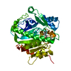
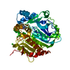
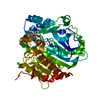
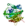
| Entry | Database: PDB / ID: 3f98 | ||||||
|---|---|---|---|---|---|---|---|
| Title | Crystal structure of human plasma platelet activating factor acetylhydrolase covalently inhibited by tabun | ||||||
 Components Components | Platelet-activating factor acetylhydrolase | ||||||
 Keywords Keywords | HYDROLASE / PLASMA PLATELET-ACTIVATING FACTOR ACETYLHYDROLASE / SECRETED PROTEIN / ALPHA/BETA-HYDROLASE-FOLD / LDL-BOUND / LIPOPROTEIN ASSOCIATED PHOSPHOLIPASE A2 / LP-PLA2 / GROUP VIIA PLA2 / GLYCOPROTEIN / LIPID DEGRADATION / POLYMORPHISM / TABUN / Disease mutation / Secreted | ||||||
| Function / homology |  Function and homology information Function and homology informationplasma lipoprotein particle oxidation / : / 1-alkyl-2-acetylglycerophosphocholine esterase / 1-alkyl-2-acetylglycerophosphocholine esterase activity / platelet activating factor metabolic process / platelet activating factor catabolic process / lipid oxidation / low-density lipoprotein particle / high-density lipoprotein particle / low-density lipoprotein particle remodeling ...plasma lipoprotein particle oxidation / : / 1-alkyl-2-acetylglycerophosphocholine esterase / 1-alkyl-2-acetylglycerophosphocholine esterase activity / platelet activating factor metabolic process / platelet activating factor catabolic process / lipid oxidation / low-density lipoprotein particle / high-density lipoprotein particle / low-density lipoprotein particle remodeling / phosphatidylcholine catabolic process / positive regulation of monocyte chemotaxis / peptide hormone processing / hydrolase activity, acting on ester bonds / Synthesis, secretion, and deacylation of Ghrelin / phospholipid binding / positive regulation of inflammatory response / extracellular region Similarity search - Function | ||||||
| Biological species |  Homo sapiens (human) Homo sapiens (human) | ||||||
| Method |  X-RAY DIFFRACTION / SYNCHROTRON / MOLECULAR REPLACEMENT / Resolution: 1.7 Å X-RAY DIFFRACTION / SYNCHROTRON / MOLECULAR REPLACEMENT / Resolution: 1.7 Å | ||||||
 Authors Authors | Samanta, U. / Bahnson, B.J. | ||||||
 Citation Citation | Journal: Biochem Pharmacol / Year: 2009 Title: Crystal structures of human group-VIIA phospholipase A2 inhibited by organophosphorus nerve agents exhibit non-aged complexes. Authors: Samanta, U. / Kirby, S.D. / Srinivasan, P. / Cerasoli, D.M. / Bahnson, B.J. | ||||||
| History |
|
- Structure visualization
| Structure viewer | Molecule: MolmilJmol/JSmol |
|---|
- Downloads & links
-Download
| PDBx/mmCIF format | 3f98.cif.gz | 277.5 KB | Display | PDBx/mmCIF format |
|---|---|---|---|---|
| PDB format | pdb3f98.ent.gz | 222.6 KB | Display | PDB format |
| PDBx/mmJSON format | 3f98.json.gz | Tree view | PDBx/mmJSON format | |
| Others |  Other downloads Other downloads |
-Validation report
| Arichive directory | https://data.pdbj.org/pub/pdb/validation_reports/f9/3f98ftp://data.pdbj.org/pub/pdb/validation_reports/f9/3f98 | HTTPS FTP |
|---|
-Related structure data
| Related structure data |  3f96C  3f97C  3f9cC  3d59S S: Starting model for refinement C: citing same article ( |
|---|---|
| Similar structure data |


-Links
 PDBj
PDBj


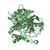
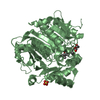
- Assembly
| Deposited unit | 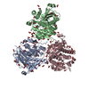
| ||||||||
|---|---|---|---|---|---|---|---|---|---|
| 1 | 
| ||||||||
| 2 | 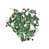
| ||||||||
| 3 | 
| ||||||||
| Unit cell |
| ||||||||
| Components on special symmetry positions |
|
-Components
| #1: Protein | Mass: 43494.367 Da / Num. of mol.: 3 / Fragment: UNP RESIDUES 47-429 Source method: isolated from a genetically manipulated source Source: (gene. exp.) Homo sapiens (human) / Gene: PLA2G7, PAFAH / Production host:  References: UniProt: Q13093, 1-alkyl-2-acetylglycerophosphocholine esterase #2: Chemical | 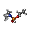  Mass: 137.117 Da / Num. of mol.: 3 / Source method: obtained synthetically / Formula: C4H12NO2P Mass: 137.117 Da / Num. of mol.: 3 / Source method: obtained synthetically / Formula: C4H12NO2P#3: Chemical | ChemComp-FMT /   Mass: 46.025 Da / Num. of mol.: 86 / Source method: obtained synthetically / Formula: CH2O2 Mass: 46.025 Da / Num. of mol.: 86 / Source method: obtained synthetically / Formula: CH2O2#4: Water | ChemComp-HOH / |  Mass: 18.015 Da / Num. of mol.: 1084 / Source method: isolated from a natural source / Formula: H2O Mass: 18.015 Da / Num. of mol.: 1084 / Source method: isolated from a natural source / Formula: H2OHas protein modification | Y | |
|---|
-Experimental details
-Experiment
| Experiment | Method: X-RAY DIFFRACTION / Number of used crystals: 1 |
|---|
- Sample preparation
| Crystal | Density Matthews: 2.52 Å3/Da / Density % sol: 51.28 % |
|---|---|
| Crystal grow | Temperature: 293 K / Method: vapor diffusion, hanging drop / pH: 7 Details: PH 7.0, VAPOR DIFFUSION, HANGING DROP, TEMPERATURE 293K, temperature 293.0K |
-Data collection
| Diffraction | Mean temperature: 100 K |
|---|---|
| Diffraction source | Source: SYNCHROTRON / Site: APS  / Beamline: 19-ID / Wavelength: 1.0055 / Wavelength: 1.0055 Å / Beamline: 19-ID / Wavelength: 1.0055 / Wavelength: 1.0055 Å |
| Detector | Type: ADSC QUANTUM 315 / Detector: CCD / Date: Aug 12, 2006 / Details: ROSENBAUM-ROCK |
| Radiation | Monochromator: SI(111) 0.990 / Protocol: SINGLE WAVELENGTH / Monochromatic (M) / Laue (L): M / Scattering type: x-ray |
| Radiation wavelength | Wavelength: 1.0055 Å / Relative weight: 1 |
| Reflection | Resolution: 1.7→50 Å / Num. all: 141507 / Num. obs: 141507 / % possible obs: 98.8 % / Observed criterion σ(F): 1 / Observed criterion σ(I): 1 / Redundancy: 10 % / Rmerge(I) obs: 0.07 / Net I/σ(I): 35.025 |
| Reflection shell | Resolution: 1.7→1.76 Å / Redundancy: 10.1 % / Rmerge(I) obs: 0.435 / Mean I/σ(I) obs: 5.59 / % possible all: 97.8 |
- Processing
| Software |
| ||||||||||||||||||||||||||||||||||||||||||||||||||||||||||||||||||||||||||||||||||||||||||
|---|---|---|---|---|---|---|---|---|---|---|---|---|---|---|---|---|---|---|---|---|---|---|---|---|---|---|---|---|---|---|---|---|---|---|---|---|---|---|---|---|---|---|---|---|---|---|---|---|---|---|---|---|---|---|---|---|---|---|---|---|---|---|---|---|---|---|---|---|---|---|---|---|---|---|---|---|---|---|---|---|---|---|---|---|---|---|---|---|---|---|---|
| Refinement | Method to determine structure: MOLECULAR REPLACEMENT Starting model: NATIVE STRUCTURE, PDB ENTRY 3D59 Resolution: 1.7→50 Å / Cor.coef. Fo:Fc: 0.953 / Cor.coef. Fo:Fc free: 0.947 / SU B: 1.692 / SU ML: 0.058 / Cross valid method: THROUGHOUT / σ(F): 1 / ESU R: 0.106 / ESU R Free: 0.099 / Stereochemistry target values: MAXIMUM LIKELIHOOD / Details: HYDROGENS HAVE BEEN ADDED IN THE RIDING POSITIONS
| ||||||||||||||||||||||||||||||||||||||||||||||||||||||||||||||||||||||||||||||||||||||||||
| Solvent computation | Ion probe radii: 0.8 Å / Shrinkage radii: 0.8 Å / VDW probe radii: 1.2 Å / Solvent model: MASK | ||||||||||||||||||||||||||||||||||||||||||||||||||||||||||||||||||||||||||||||||||||||||||
| Displacement parameters | Biso mean: 20.122 Å2
| ||||||||||||||||||||||||||||||||||||||||||||||||||||||||||||||||||||||||||||||||||||||||||
| Refinement step | Cycle: LAST / Resolution: 1.7→50 Å
| ||||||||||||||||||||||||||||||||||||||||||||||||||||||||||||||||||||||||||||||||||||||||||
| Refine LS restraints |
| ||||||||||||||||||||||||||||||||||||||||||||||||||||||||||||||||||||||||||||||||||||||||||
| LS refinement shell | Resolution: 1.7→1.744 Å / Total num. of bins used: 20
|
