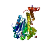
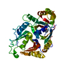
[English] 日本語
Yorodumi- PDB-6nje: Crystal structure of the motor domain of human kinesin family mem... -
+Open data
-Basic information
Entry | Database: PDB / ID: 6nje | |||||||||
---|---|---|---|---|---|---|---|---|---|---|
Title | Crystal structure of the motor domain of human kinesin family member 22 | |||||||||
Components | Kinesin-like protein KIF22 | |||||||||
Keywords | TRANSPORT PROTEIN / limited proteolysis / kinesin / structural genomics consortium / motor domain / adp / SGC | |||||||||
Function / homology | Function and homology information metaphase chromosome alignment / Kinesins / sister chromatid cohesion / COPI-dependent Golgi-to-ER retrograde traffic / kinesin complex / microtubule motor activity / mitotic metaphase chromosome alignment / microtubule-based movement / MHC class II antigen presentation / mitotic spindle ...metaphase chromosome alignment / Kinesins / sister chromatid cohesion / COPI-dependent Golgi-to-ER retrograde traffic / kinesin complex / microtubule motor activity / mitotic metaphase chromosome alignment / microtubule-based movement / MHC class II antigen presentation / mitotic spindle / kinetochore / mitotic cell cycle / microtubule binding / microtubule / nuclear speck / DNA repair / chromatin / ATP hydrolysis activity / DNA binding / ATP binding / nucleus / cytosol Similarity search - Function | |||||||||
Biological species | Homo sapiens (human) | |||||||||
Method | X-RAY DIFFRACTION / MOLECULAR REPLACEMENT / Resolution: 2.2 Å | |||||||||
Authors | Walker, B.C. / Zhu, H. / Tempel, W. / Arrowsmith, C.H. / Edwards, A.M. / Park, H. / Cochran, J.C. / Structural Genomics Consortium (SGC) | |||||||||
Citation | Journal: To Be Published Title: Crystal structure of the motor domain of human kinesin family member 22 Authors: Walker, B.C. / Zhu, H. / Tempel, W. / Arrowsmith, C.H. / Edwards, A.M. / Park, H. / Structural Genomics Consortium (SGC) | |||||||||
History |
|
-Structure visualization
Structure viewer | Molecule: MolmilJmol/JSmol |
---|
-Downloads & links
-Download
PDBx/mmCIF format | 6nje.cif.gz | 81.3 KB | Display | PDBx/mmCIF format |
---|---|---|---|---|
PDB format | pdb6nje.ent.gz | 56.2 KB | Display | PDB format |
PDBx/mmJSON format | 6nje.json.gz | Tree view | PDBx/mmJSON format | |
Others | Other downloads |
-Validation report
Arichive directory | https://data.pdbj.org/pub/pdb/validation_reports/nj/6nje ftp://data.pdbj.org/pub/pdb/validation_reports/nj/6nje | HTTPS FTP |
---|
-Related structure data
Related structure data | 3b6uS 3bfn S: Starting model for refinement |
---|---|
Similar structure data | |
Experimental dataset #1 | Data reference: 10.5281/zenodo.1455747 / Data set type: diffraction image data |
-Links
-Assembly
Deposited unit |
| ||||||||
---|---|---|---|---|---|---|---|---|---|
1 |
| ||||||||
Unit cell |
|
-Components
-Protein , 1 types, 1 molecules A
#1: Protein | Mass: 42132.816 Da / Num. of mol.: 1 Source method: isolated from a genetically manipulated source Source: (gene. exp.) Homo sapiens (human) / Gene: KIF22, KID, KNSL4 / Plasmid: pET28-MHL / Production host: Escherichia coli (E. coli) / Strain (production host): BL21 (DE3) codon(+) RIL / References: UniProt: Q14807 |
---|
-Non-polymers , 5 types, 96 molecules
#2: Chemical | ChemComp-MG / | ||||
---|---|---|---|---|---|
#3: Chemical | ChemComp-ADP / | ||||
#4: Chemical | #5: Chemical | ChemComp-NA / | #6: Water | ChemComp-HOH / | |
-Experimental details
-Experiment
Experiment | Method: X-RAY DIFFRACTION / Number of used crystals: 1 |
---|
-Sample preparation
Crystal | Density Matthews: 1.81 Å3/Da / Density % sol: 31.94 % |
---|---|
Crystal grow | Temperature: 291 K / Method: vapor diffusion / pH: 8.5 / Details: 3.2 M NaCl, 0.1 M Tris |
-Data collection
Diffraction | Mean temperature: 100 K / Serial crystal experiment: N | |||||||||||||||||||||||||||||||||
---|---|---|---|---|---|---|---|---|---|---|---|---|---|---|---|---|---|---|---|---|---|---|---|---|---|---|---|---|---|---|---|---|---|---|
Diffraction source | Source: ROTATING ANODE / Type: RIGAKU FR-E / Wavelength: 1.5418 Å | |||||||||||||||||||||||||||||||||
Detector | Type: RIGAKU RAXIS / Detector: IMAGE PLATE / Date: Oct 10, 2007 | |||||||||||||||||||||||||||||||||
Radiation | Protocol: SINGLE WAVELENGTH / Monochromatic (M) / Laue (L): M / Scattering type: x-ray | |||||||||||||||||||||||||||||||||
Radiation wavelength | Wavelength: 1.5418 Å / Relative weight: 1 | |||||||||||||||||||||||||||||||||
Reflection | Resolution: 2.2→35.48 Å / Num. obs: 16157 / % possible obs: 99.5 % / Redundancy: 6.8 % / Biso Wilson estimate: 39.71 Å2 / CC1/2: 0.998 / Rmerge(I) obs: 0.121 / Rpim(I) all: 0.049 / Rrim(I) all: 0.13 / Net I/σ(I): 16.1 / Num. measured all: 109199 / Scaling rejects: 15 | |||||||||||||||||||||||||||||||||
Reflection shell | Diffraction-ID: 1
|
-Processing
Software |
| ||||||||||||||||||||||||||||||||||||||||||||||||||||||||||||||||||||||||||||||||||||||||||||||||||||||||||||
---|---|---|---|---|---|---|---|---|---|---|---|---|---|---|---|---|---|---|---|---|---|---|---|---|---|---|---|---|---|---|---|---|---|---|---|---|---|---|---|---|---|---|---|---|---|---|---|---|---|---|---|---|---|---|---|---|---|---|---|---|---|---|---|---|---|---|---|---|---|---|---|---|---|---|---|---|---|---|---|---|---|---|---|---|---|---|---|---|---|---|---|---|---|---|---|---|---|---|---|---|---|---|---|---|---|---|---|---|---|
Refinement | Method to determine structure: MOLECULAR REPLACEMENT Starting model: continued refinement of model from PDB entry 3BFN. Previously solved using coordinates from PDB entry 3B6U. Resolution: 2.2→35.48 Å / Cor.coef. Fo:Fc: 0.929 / Cor.coef. Fo:Fc free: 0.891 / Rfactor Rfree error: 0 / SU R Cruickshank DPI: 0.276 / Cross valid method: FREE R-VALUE / σ(F): 0 / SU R Blow DPI: 0.288 / SU Rfree Blow DPI: 0.223 / SU Rfree Cruickshank DPI: 0.222 Details: Discontinuous electron density suggests that some residues between positions 55 and 71, which have been omitted from the model, form an additional strand adjacent to the strand that includes ...Details: Discontinuous electron density suggests that some residues between positions 55 and 71, which have been omitted from the model, form an additional strand adjacent to the strand that includes residues 74 through 76.
| ||||||||||||||||||||||||||||||||||||||||||||||||||||||||||||||||||||||||||||||||||||||||||||||||||||||||||||
Displacement parameters | Biso max: 128.33 Å2 / Biso mean: 39.38 Å2 / Biso min: 15.37 Å2
| ||||||||||||||||||||||||||||||||||||||||||||||||||||||||||||||||||||||||||||||||||||||||||||||||||||||||||||
Refine analyze | Luzzati coordinate error obs: 0.28 Å | ||||||||||||||||||||||||||||||||||||||||||||||||||||||||||||||||||||||||||||||||||||||||||||||||||||||||||||
Refinement step | Cycle: final / Resolution: 2.2→35.48 Å
| ||||||||||||||||||||||||||||||||||||||||||||||||||||||||||||||||||||||||||||||||||||||||||||||||||||||||||||
Refine LS restraints |
| ||||||||||||||||||||||||||||||||||||||||||||||||||||||||||||||||||||||||||||||||||||||||||||||||||||||||||||
LS refinement shell | Resolution: 2.2→2.35 Å / Total num. of bins used: 8
|