 Movie
Movie Controller


+ Open data

- Basic information
| Entry | Database: PDB / ID: 4bn2 | |||||||||
|---|---|---|---|---|---|---|---|---|---|---|
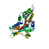
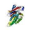
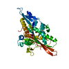
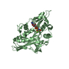
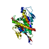
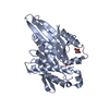
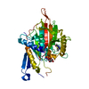
| Title | The crystal structure of kinesin-like protein KIF15 | |||||||||
 Components Components | KINESIN-LIKE PROTEIN KIF15 | |||||||||
 Keywords Keywords | MOTOR PROTEIN / KINESIN / MOTOR DOMAIN | |||||||||
| Function / homology |  Function and homology information Function and homology informationplus-end kinesin complex / centrosome separation / plus-end-directed microtubule motor activity / Kinesins / microtubule motor activity / kinesin complex / COPI-dependent Golgi-to-ER retrograde traffic / microtubule-based movement / cytoskeletal motor activity / mitotic spindle assembly ...plus-end kinesin complex / centrosome separation / plus-end-directed microtubule motor activity / Kinesins / microtubule motor activity / kinesin complex / COPI-dependent Golgi-to-ER retrograde traffic / microtubule-based movement / cytoskeletal motor activity / mitotic spindle assembly / MHC class II antigen presentation / spindle pole / mitotic cell cycle / microtubule binding / microtubule / centrosome / ATP hydrolysis activity / ATP binding / membrane / cytoplasm / cytosol Similarity search - Function | |||||||||
| Biological species |  HOMO SAPIENS (human) HOMO SAPIENS (human) | |||||||||
| Method |  X-RAY DIFFRACTION / SYNCHROTRON / MOLECULAR REPLACEMENT / Resolution: 2.695 Å X-RAY DIFFRACTION / SYNCHROTRON / MOLECULAR REPLACEMENT / Resolution: 2.695 Å | |||||||||
 Authors Authors | Klejnot, M. / Falnikar, A. / Ulaganathan, V. / Cross, R. / Baas, P. / Kozielski, F. | |||||||||
 Citation Citation | Journal: Acta Crystallogr.,Sect.D / Year: 2014 Title: The Crystal Structure and Biochemical Characterization of Kif15: A Bifunctional Molecular Motor Involved in Bipolar Spindle Formation and Neuronal Development Authors: Klejnot, M. / Falnikar, A. / Ulaganathan, V. / Cross, R.A. / Baas, P.W. / Kozielski, F. | |||||||||
| History |
|
- Structure visualization
| Structure viewer | Molecule: MolmilJmol/JSmol |
|---|
- Downloads & links
-Download
| PDBx/mmCIF format | 4bn2.cif.gz | 204.3 KB | Display | PDBx/mmCIF format |
|---|---|---|---|---|
| PDB format | pdb4bn2.ent.gz | 160.5 KB | Display | PDB format |
| PDBx/mmJSON format | 4bn2.json.gz | Tree view | PDBx/mmJSON format | |
| Others |  Other downloads Other downloads |
-Validation report
| Arichive directory | https://data.pdbj.org/pub/pdb/validation_reports/bn/4bn2ftp://data.pdbj.org/pub/pdb/validation_reports/bn/4bn2 | HTTPS FTP |
|---|
-Related structure data
| Related structure data | 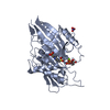 1t5cS S: Starting model for refinement |
|---|---|
| Similar structure data |


-Links
 PDBj
PDBj


- Assembly
| Deposited unit | 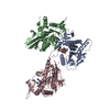
| ||||||||||||||||||||||||||||||||||||
|---|---|---|---|---|---|---|---|---|---|---|---|---|---|---|---|---|---|---|---|---|---|---|---|---|---|---|---|---|---|---|---|---|---|---|---|---|---|
| 1 | 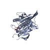
| ||||||||||||||||||||||||||||||||||||
| 2 | 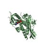
| ||||||||||||||||||||||||||||||||||||
| 3 | 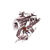
| ||||||||||||||||||||||||||||||||||||
| Unit cell |
| ||||||||||||||||||||||||||||||||||||
| Components on special symmetry positions |
| ||||||||||||||||||||||||||||||||||||
| Noncrystallographic symmetry (NCS) | NCS domain:
NCS domain segments:
NCS oper:
|
-Components
| #1: Protein | Mass: 39374.340 Da / Num. of mol.: 3 / Fragment: MOTOR DOMAIN, RESIDUES 19-375 Source method: isolated from a genetically manipulated source Source: (gene. exp.) HOMO SAPIENS (human) / Plasmid: PETM20MOD / Production host:  #2: Chemical |   Mass: 427.201 Da / Num. of mol.: 3 / Source method: obtained synthetically / Formula: C10H15N5O10P2 / Comment: ADP, energy-carrying molecule*YM Mass: 427.201 Da / Num. of mol.: 3 / Source method: obtained synthetically / Formula: C10H15N5O10P2 / Comment: ADP, energy-carrying molecule*YM#3: Chemical |   Mass: 24.305 Da / Num. of mol.: 3 / Source method: obtained synthetically / Formula: Mg Mass: 24.305 Da / Num. of mol.: 3 / Source method: obtained synthetically / Formula: Mg#4: Water | ChemComp-HOH / |  Mass: 18.015 Da / Num. of mol.: 203 / Source method: isolated from a natural source / Formula: H2O Mass: 18.015 Da / Num. of mol.: 203 / Source method: isolated from a natural source / Formula: H2OHas protein modification | Y | Sequence details | RESIDUES 1 TO 4 CHANGED DUE TO CLONING | |
|---|
-Experimental details
-Experiment
| Experiment | Method: X-RAY DIFFRACTION / Number of used crystals: 1 |
|---|
- Sample preparation
| Crystal | Density Matthews: 2.74 Å3/Da / Density % sol: 57.22 % / Description: NONE |
|---|---|
| Crystal grow | Temperature: 292 K / pH: 6 Details: 25% POLYETHYLENE GLYCOL-3350 0.20 M MAGNESIUM CHLORIDE 0.1 M TRIS-HC; PH 8.5 |
-Data collection
| Diffraction | Mean temperature: 93 K |
|---|---|
| Diffraction source | Source: SYNCHROTRON / Site: ESRF  / Beamline: ID23-1 / Wavelength: 0.8726 / Beamline: ID23-1 / Wavelength: 0.8726 |
| Detector | Type: ADSC QUANTUM 315r / Detector: CCD / Date: Nov 23, 2009 |
| Radiation | Protocol: SINGLE WAVELENGTH / Monochromatic (M) / Laue (L): M / Scattering type: x-ray |
| Radiation wavelength | Wavelength: 0.8726 Å / Relative weight: 1 |
| Reflection | Resolution: 2.7→30 Å / Num. obs: 29592 / % possible obs: 89.7 % / Observed criterion σ(I): 2 / Redundancy: 3.7 % / Biso Wilson estimate: 37.87 Å2 / Rmerge(I) obs: 0.098 / Net I/σ(I): 8.6 |
| Reflection shell | Highest resolution: 2.7 Å / Redundancy: 3.5 % / Rmerge(I) obs: 0.378 / Mean I/σ(I) obs: 3.1 / % possible all: 86.5 |
- Processing
| Software |
| ||||||||||||||||||||||||||||||||||||||||||
|---|---|---|---|---|---|---|---|---|---|---|---|---|---|---|---|---|---|---|---|---|---|---|---|---|---|---|---|---|---|---|---|---|---|---|---|---|---|---|---|---|---|---|---|
| Refinement | Method to determine structure: MOLECULAR REPLACEMENT Starting model: 1T5C Resolution: 2.695→29.486 Å / SU ML: 0.88 / σ(F): 1.34 / Phase error: 27.16 / Stereochemistry target values: ML
| ||||||||||||||||||||||||||||||||||||||||||
| Solvent computation | Shrinkage radii: 0.95 Å / VDW probe radii: 1.2 Å / Solvent model: FLAT BULK SOLVENT MODEL / Bsol: 28.074 Å2 / ksol: 0.335 e/Å3 | ||||||||||||||||||||||||||||||||||||||||||
| Displacement parameters |
| ||||||||||||||||||||||||||||||||||||||||||
| Refinement step | Cycle: LAST / Resolution: 2.695→29.486 Å
| ||||||||||||||||||||||||||||||||||||||||||
| Refine LS restraints |
| ||||||||||||||||||||||||||||||||||||||||||
| Refine LS restraints NCS |
| ||||||||||||||||||||||||||||||||||||||||||
| LS refinement shell |
|
