- PDB-6ncm: Crystal structure of the human FOXN3 DNA binding domain in comple... -
+
Open data
ID or keywords:
Loading...
-
Basic information
Entry
Database: PDB / ID: 6ncm
Title
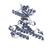
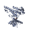
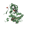
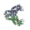
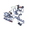
Crystal structure of the human FOXN3 DNA binding domain in complex with a forkhead-like (FHL) DNA sequence
Components
DNA (5'-D(*AP*TP*AP*GP*CP*GP*TP*CP*TP*TP*AP*GP*CP*AP*TP*G)-3')
DNA (5'-D(*TP*CP*AP*TP*GP*CP*TP*AP*AP*GP*AP*CP*GP*CP*TP*A)-3')
Forkhead box protein N3FOX proteins
Keywords
DNA BINDING PROTEIN/DNA / sequence specific DNA binding / regulation of transcription DNA templated / DNA Binding Transcription Factor Activity / forkhead / DNA BINDING PROTEIN / DNA BINDING PROTEIN-DNA complex
Function / homology
Function and homology information
craniofacial suture morphogenesis / : / mitotic G2 DNA damage checkpoint signaling / cis-regulatory region sequence-specific DNA binding / DNA-binding transcription factor activity, RNA polymerase II-specific / DNA-binding transcription factor activity / negative regulation of DNA-templated transcription / chromatin / regulation of DNA-templated transcription / nucleus Similarity search - Function
Fork head domain conserved site1 / Fork head domain signature 1. / Fork head domain / Forkhead domain / Fork head domain profile. / FORKHEAD / Fork head domain conserved site 2 / Fork head domain signature 2. / Winged helix-like DNA-binding domain superfamily/Winged helix DNA-binding domain / Arc Repressor Mutant, subunit A ...Fork head domain conserved site1 / Fork head domain signature 1. / Fork head domain / Forkhead domain / Fork head domain profile. / FORKHEAD / Fork head domain conserved site 2 / Fork head domain signature 2. / Winged helix-like DNA-binding domain superfamily/Winged helix DNA-binding domain / Arc Repressor Mutant, subunit A / Winged helix DNA-binding domain superfamily / Winged helix-like DNA-binding domain superfamily / Orthogonal Bundle / Mainly Alpha Similarity search - Domain/homology
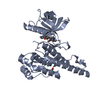
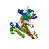
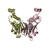
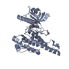
A: Forkhead box protein N3 C: DNA (5'-D(*AP*TP*AP*GP*CP*GP*TP*CP*TP*TP*AP*GP*CP*AP*TP*G)-3') D: DNA (5'-D(*TP*CP*AP*TP*GP*CP*TP*AP*AP*GP*AP*CP*GP*CP*TP*A)-3') B: Forkhead box protein N3 hetero molecules
In the structure databanks used in Yorodumi, some data are registered as the other names, "COVID-19 virus" and "2019-nCoV". Here are the details of the virus and the list of structure data.
Jan 31, 2019. EMDB accession codes are about to change! (news from PDBe EMDB page)
EMDB accession codes are about to change! (news from PDBe EMDB page)
The allocation of 4 digits for EMDB accession codes will soon come to an end. Whilst these codes will remain in use, new EMDB accession codes will include an additional digit and will expand incrementally as the available range of codes is exhausted. The current 4-digit format prefixed with “EMD-” (i.e. EMD-XXXX) will advance to a 5-digit format (i.e. EMD-XXXXX), and so on. It is currently estimated that the 4-digit codes will be depleted around Spring 2019, at which point the 5-digit format will come into force.
The EM Navigator/Yorodumi systems omit the EMD- prefix.
Related info.:Q: What is EMD? / ID/Accession-code notation in Yorodumi/EM Navigator
Yorodumi is a browser for structure data from EMDB, PDB, SASBDB, etc.
This page is also the successor to EM Navigator detail page, and also detail information page/front-end page for Omokage search.
The word "yorodu" (or yorozu) is an old Japanese word meaning "ten thousand". "mi" (miru) is to see.
Related info.:EMDB / PDB / SASBDB / Comparison of 3 databanks / Yorodumi Search / Aug 31, 2016. New EM Navigator & Yorodumi / Yorodumi Papers / Jmol/JSmol / Function and homology information / Changes in new EM Navigator and Yorodumi
 Movie
Movie Controller

 Yorodumi
 Open data
 Basic information
 Components
 Keywords
 forkhead /
  Function and homology information
 Authors
 United States, 2items
  Citation
 Structure visualization
 Downloads & links
 Other downloads

 PDBj
PDBj


 Assembly
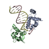


 Mass: 24.305 Da / Num. of mol.: 2 / Source method: obtained synthetically / Formula: Mg
 Mass: 18.015 Da / Num. of mol.: 13 / Source method: isolated from a natural source / Formula: H2O
 Sample preparation
 Processing