Mass: 18.015 Da / Num. of mol.: 118 / Source method: isolated from a natural source / Formula: H2O
Has protein modification
N
Nonpolymer details
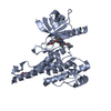
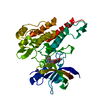
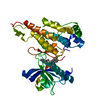
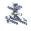
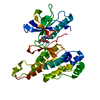
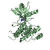
TYR1054 AND TYR1059 WERE PHOSPHORYLATED BY AN AUTOPHOSPHORYLATION REACTION RUN ON THE PROTEIN PRIOR ...TYR1054 AND TYR1059 WERE PHOSPHORYLATED BY AN AUTOPHOSPHORYLATION REACTION RUN ON THE PROTEIN PRIOR TO CRYSTALLIZATION. THE PHOSPHORYLATED TYR IS REPRESENTED BY PTR, PHOSPHONOTYROSINE, PTR1054 AND PTR1059. THESE RESIDUES ARE PART OF THE KINASE ACTIVATION LOOP AND THEY ARE DISORDERED IN THE STRUCTURE AND CONSEQUENTLY NOT SEEN IN THE ELECTRON DENSITY.
-
Experimental details
-
Experiment
Experiment
Method: X-RAY DIFFRACTION / Number of used crystals: 1
-
Sample preparation
Crystal
Density Matthews: 2.29 Å3/Da / Density % sol: 46.25 %
Resolution: 2.5→2.59 Å / Redundancy: 4.1 % / Rmerge(I) obs: 0.494 / Mean I/σ(I) obs: 3.1 / Num. unique all: 2249 / Χ2: 1.075 / % possible all: 100
-
Processing
Software
Name
Version
Classification
NB
SCALEPACK
datascaling
CNS
refinement
PDB_EXTRACT
1.401
dataextraction
DENZO
datareduction
CNS
phasing
Refinement
Method to determine structure: FOURIER SYNTHESIS / Resolution: 2.5→30 Å / Data cutoff high absF: 10000 / Data cutoff low absF: 0 / Cross valid method: THROUGHOUT / σ(F): 0
Rfactor
Num. reflection
% reflection
Selection details
Rfree
0.263
1341
5.9 %
random
Rwork
0.211
-
-
-
all
-
22857
-
-
obs
-
22848
100 %
-
Displacement parameters
Baniso -1
Baniso -2
Baniso -3
1-
4.569 Å2
0 Å2
-3.871 Å2
2-
-
4.238 Å2
0 Å2
3-
-
-
-8.807 Å2
Refine analyze
Free
Obs
Luzzati coordinate error
0.4 Å
0.3 Å
Luzzati d res low
-
5 Å
Luzzati sigma a
0.37 Å
0.29 Å
Refinement step
Cycle: LAST / Resolution: 2.5→30 Å
Protein
Nucleic acid
Ligand
Solvent
Total
Num. atoms
4491
0
60
118
4669
Refine LS restraints
Refine-ID
Type
Dev ideal
X-RAY DIFFRACTION
c_angle_deg
1.27
X-RAY DIFFRACTION
c_bond_d
0.007
X-RAY DIFFRACTION
c_dihedral_angle_d
21.3
X-RAY DIFFRACTION
c_improper_angle_d
0.76
+
About Yorodumi
-
News
-
Feb 9, 2022. New format data for meta-information of EMDB entries
New format data for meta-information of EMDB entries
Version 3 of the EMDB header file is now the official format.
The previous official version 1.9 will be removed from the archive.
In the structure databanks used in Yorodumi, some data are registered as the other names, "COVID-19 virus" and "2019-nCoV". Here are the details of the virus and the list of structure data.
Jan 31, 2019. EMDB accession codes are about to change! (news from PDBe EMDB page)
EMDB accession codes are about to change! (news from PDBe EMDB page)
The allocation of 4 digits for EMDB accession codes will soon come to an end. Whilst these codes will remain in use, new EMDB accession codes will include an additional digit and will expand incrementally as the available range of codes is exhausted. The current 4-digit format prefixed with “EMD-” (i.e. EMD-XXXX) will advance to a 5-digit format (i.e. EMD-XXXXX), and so on. It is currently estimated that the 4-digit codes will be depleted around Spring 2019, at which point the 5-digit format will come into force.
The EM Navigator/Yorodumi systems omit the EMD- prefix.
Related info.:Q: What is EMD? / ID/Accession-code notation in Yorodumi/EM Navigator
Yorodumi is a browser for structure data from EMDB, PDB, SASBDB, etc.
This page is also the successor to EM Navigator detail page, and also detail information page/front-end page for Omokage search.
The word "yorodu" (or yorozu) is an old Japanese word meaning "ten thousand". "mi" (miru) is to see.
Related info.:EMDB / PDB / SASBDB / Comparison of 3 databanks / Yorodumi Search / Aug 31, 2016. New EM Navigator & Yorodumi / Yorodumi Papers / Jmol/JSmol / Function and homology information / Changes in new EM Navigator and Yorodumi
 Movie
Movie Controller

 Yorodumi
 Open data
 Basic information
 Components
 Keywords
 Function and homology information
 Homo sapiens (human)
 X-RAY DIFFRACTION /
  Authors
 Citation
 Structure visualization
 Downloads & links
 Other downloads


 PDBj
PDBj


 Assembly
 Trichoplusia ni (cabbage looper)
 Mass: 397.472 Da / Num. of mol.: 2 / Source method: obtained synthetically / Formula: C24H23N5O
 Mass: 18.015 Da / Num. of mol.: 118 / Source method: isolated from a natural source / Formula: H2O
 Sample preparation
 / Beamline: 31-ID / Wavelength: 0.9793 Å
 Processing