 Movie
Movie Controller

[English] 日本語
 Yorodumi
- PDB-6n2o: 2-oxoglutarate:ferredoxin oxidoreductase from Magnetococcus marin... -
+ Open data

- Basic information
| Entry | Database: PDB / ID: 6n2o | |||||||||
|---|---|---|---|---|---|---|---|---|---|---|
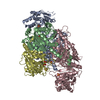
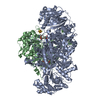
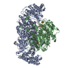
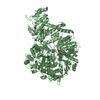
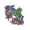
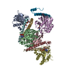
| Title | 2-oxoglutarate:ferredoxin oxidoreductase from Magnetococcus marinus with 2-oxoglutarate, coenzyme A and succinyl-CoA bound | |||||||||
 Components Components |
| |||||||||
 Keywords Keywords |  OXIDOREDUCTASE / thiamine pyrophosphate / [4Fe-4S] cluster / carbon fixation / reductive tricarboxylic acid cycle / rTCA / 2-oxoglutarate:ferredoxin oxidoreductase / electron transfer OXIDOREDUCTASE / thiamine pyrophosphate / [4Fe-4S] cluster / carbon fixation / reductive tricarboxylic acid cycle / rTCA / 2-oxoglutarate:ferredoxin oxidoreductase / electron transfer | |||||||||
| Function / homology |  Function and homology information Function and homology informationoxidoreductase activity, acting on the aldehyde or oxo group of donors / thiamine pyrophosphate binding / catalytic activitySimilarity search - Function | |||||||||
| Biological species |  Magnetococcus marinus (bacteria) Magnetococcus marinus (bacteria) | |||||||||
| Method | X-RAY DIFFRACTION / SYNCHROTRON / MOLECULAR REPLACEMENT / Resolution: 2.824 Å | |||||||||
 Authors Authors | Chen, P.Y.-T. / Drennan, C.L. | |||||||||
| Funding support |  United States, 2items United States, 2items
| |||||||||
 Citation Citation | Journal: Joule / Year: 2019 Title: A reverse TCA cycle 2-oxoacid:ferredoxin oxidoreductase that makes C-C bonds from CO2. Authors: Chen, P.Y. / Li, B. / Drennan, C.L. / Elliott, S.J. | |||||||||
| History |
|
- Structure visualization
| Structure viewer | Molecule: MolmilJmol/JSmol |
|---|
- Downloads & links
-Download
| PDBx/mmCIF format | 6n2o.cif.gz | 341.4 KB | Display | PDBx/mmCIF format |
|---|---|---|---|---|
| PDB format | pdb6n2o.ent.gz | 268.8 KB | Display | PDB format |
| PDBx/mmJSON format | 6n2o.json.gz | Tree view | PDBx/mmJSON format | |
| Others |  Other downloads Other downloads |
-Validation report
| Arichive directory | https://data.pdbj.org/pub/pdb/validation_reports/n2/6n2oftp://data.pdbj.org/pub/pdb/validation_reports/n2/6n2o | HTTPS FTP |
|---|
-Related structure data
| Related structure data |  6n2nSC S: Starting model for refinement C: citing same article ( |
|---|---|
| Similar structure data |

-Links
 PDBj
PDBj


- Assembly
| Deposited unit | 
| ||||||||
|---|---|---|---|---|---|---|---|---|---|
| 1 |
| ||||||||
| Unit cell |
|
-Components
-Protein , 2 types, 4 molecules ACBD
| #1: Protein | Mass: 62159.688 Da / Num. of mol.: 2 Source method: isolated from a genetically manipulated source Source: (gene. exp.) Magnetococcus marinus (strain ATCC BAA-1437 / JCM 17883 / MC-1) (bacteria)Strain: ATCC BAA-1437 / JCM 17883 / MC-1 / Gene: Mmc1_1749 / Production host: Escherichia coli (E. coli) / References: UniProt: A0L8G4#2: Protein | Mass: 31706.275 Da / Num. of mol.: 2 Source method: isolated from a genetically manipulated source Source: (gene. exp.) Magnetococcus marinus (strain ATCC BAA-1437 / JCM 17883 / MC-1) (bacteria)Strain: ATCC BAA-1437 / JCM 17883 / MC-1 / Gene: Mmc1_1750 / Production host: Escherichia coli (E. coli) / References: UniProt: A0L8G5 |
|---|
-Non-polymers , 7 types, 31 molecules 


| #3: Chemical | ChemComp-AKG / Α-Ketoglutaric acid Mass: 146.098 Da / Num. of mol.: 1 / Source method: obtained synthetically / Formula: C5H6O5 / Feature type: SUBJECT OF INVESTIGATION Mass: 146.098 Da / Num. of mol.: 1 / Source method: obtained synthetically / Formula: C5H6O5 / Feature type: SUBJECT OF INVESTIGATION | ||||||||
|---|---|---|---|---|---|---|---|---|---|
| #4: Chemical | ChemComp-COA / Coenzyme A Mass: 767.534 Da / Num. of mol.: 1 / Source method: obtained synthetically / Formula: C21H36N7O16P3S / Feature type: SUBJECT OF INVESTIGATION Mass: 767.534 Da / Num. of mol.: 1 / Source method: obtained synthetically / Formula: C21H36N7O16P3S / Feature type: SUBJECT OF INVESTIGATION | ||||||||
| #5: Chemical | Iron–sulfur cluster Mass: 351.640 Da / Num. of mol.: 2 / Source method: obtained synthetically / Formula: Fe4S4 Mass: 351.640 Da / Num. of mol.: 2 / Source method: obtained synthetically / Formula: Fe4S4#6: Chemical | Thiamine pyrophosphate Mass: 425.314 Da / Num. of mol.: 2 / Source method: obtained synthetically / Formula: C12H19N4O7P2S Mass: 425.314 Da / Num. of mol.: 2 / Source method: obtained synthetically / Formula: C12H19N4O7P2S#7: Chemical |  Mass: 24.305 Da / Num. of mol.: 2 / Source method: obtained synthetically / Formula: Mg Mass: 24.305 Da / Num. of mol.: 2 / Source method: obtained synthetically / Formula: Mg#8: Chemical | ChemComp-SCA / | Succinyl-CoA Mass: 867.607 Da / Num. of mol.: 1 / Source method: obtained synthetically / Formula: C25H40N7O19P3S / Feature type: SUBJECT OF INVESTIGATION Mass: 867.607 Da / Num. of mol.: 1 / Source method: obtained synthetically / Formula: C25H40N7O19P3S / Feature type: SUBJECT OF INVESTIGATION#9: Water | ChemComp-HOH / | WaterMass: 18.015 Da / Num. of mol.: 22 / Source method: isolated from a natural source / Formula: H2O |
-Experimental details
-Experiment
| Experiment | Method: X-RAY DIFFRACTION / Number of used crystals: 1 |
|---|
- Sample preparation
| Crystal | Density Matthews: 2.34 Å3/Da / Density % sol: 47.38 % |
|---|---|
| Crystal grow | Temperature: 300 K / Method: vapor diffusion, sitting drop Details: 27% (w/v) PEG 4000 and 0.08 M sodium cacodylate pH 5.5 |
-Data collection
| Diffraction | Mean temperature: 100 K |
|---|---|
| Diffraction source | Source: SYNCHROTRON / Site: APS / Beamline: 24-ID-C / Wavelength: 0.9791 Å |
| Detector | Type: DECTRIS PILATUS 6M / Detector: PIXEL / Date: Nov 13, 2015 |
| Radiation | Protocol: SINGLE WAVELENGTH / Monochromatic (M) / Laue (L): M / Scattering type: x-ray |
| Radiation wavelength | Wavelength: 0.9791 Å / Relative weight: 1 |
| Reflection | Resolution: 2.8→100 Å / Num. obs: 38629 / % possible obs: 90.7 % / Redundancy: 8.1 % / Rsym value: 0.133 / Net I/σ(I): 10.9 |
| Reflection shell | Resolution: 2.8→2.9 Å / Num. unique obs: 2935 / CC1/2: 0.857 |
- Processing
| Software |
| |||||||||||||||||||||||||||||||||||||||||||||||||||||||||||||||||||||||||||||||||||||||||||||||||||||||||
|---|---|---|---|---|---|---|---|---|---|---|---|---|---|---|---|---|---|---|---|---|---|---|---|---|---|---|---|---|---|---|---|---|---|---|---|---|---|---|---|---|---|---|---|---|---|---|---|---|---|---|---|---|---|---|---|---|---|---|---|---|---|---|---|---|---|---|---|---|---|---|---|---|---|---|---|---|---|---|---|---|---|---|---|---|---|---|---|---|---|---|---|---|---|---|---|---|---|---|---|---|---|---|---|---|---|---|
| Refinement | Method to determine structure: MOLECULAR REPLACEMENT Starting model: 6N2N Resolution: 2.824→90.006 Å / SU ML: 0.37 / Cross valid method: FREE R-VALUE / σ(F): 1.35 / Phase error: 29.8
| |||||||||||||||||||||||||||||||||||||||||||||||||||||||||||||||||||||||||||||||||||||||||||||||||||||||||
| Solvent computation | Shrinkage radii: 0.9 Å / VDW probe radii: 1.11 Å | |||||||||||||||||||||||||||||||||||||||||||||||||||||||||||||||||||||||||||||||||||||||||||||||||||||||||
| Refinement step | Cycle: LAST / Resolution: 2.824→90.006 Å
| |||||||||||||||||||||||||||||||||||||||||||||||||||||||||||||||||||||||||||||||||||||||||||||||||||||||||
| Refine LS restraints |
| |||||||||||||||||||||||||||||||||||||||||||||||||||||||||||||||||||||||||||||||||||||||||||||||||||||||||
| LS refinement shell |
|
