 Movie
Movie Controller

[English] 日本語
 Yorodumi
- PDB-6dwy: Hermes transposase deletion dimer complex with (C/G) DNA and Ca2+ -
+ Open data

- Basic information
| Entry | Database: PDB / ID: 6dwy | ||||||
|---|---|---|---|---|---|---|---|
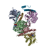
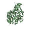
| Title | Hermes transposase deletion dimer complex with (C/G) DNA and Ca2+ | ||||||
 Components Components |
| ||||||
 Keywords Keywords | dna binding protein/dna / transposase / DNA BINDING PROTEIN / dna binding protein-dna complex | ||||||
| Function / homology |  Function and homology information Function and homology informationprotein dimerization activity / regulation of transcription by RNA polymerase II / DNA binding / zinc ion binding / nucleus Similarity search - Function | ||||||
| Biological species |  Musca domestica (house fly) Musca domestica (house fly) | ||||||
| Method |  X-RAY DIFFRACTION / SYNCHROTRON / MOLECULAR REPLACEMENT / Resolution: 3.2 Å X-RAY DIFFRACTION / SYNCHROTRON / MOLECULAR REPLACEMENT / Resolution: 3.2 Å | ||||||
 Authors Authors | Dyda, F. / Hickman, A.B. | ||||||
 Citation Citation | Journal: Nucleic Acids Res. / Year: 2018 Title: Structural insights into the mechanism of double strand break formation by Hermes, a hAT family eukaryotic DNA transposase. Authors: Hickman, A.B. / Voth, A.R. / Ewis, H. / Li, X. / Craig, N.L. / Dyda, F. | ||||||
| History |
|
- Structure visualization
| Structure viewer | Molecule: MolmilJmol/JSmol |
|---|
- Downloads & links
-Download
| PDBx/mmCIF format | 6dwy.cif.gz | 140.4 KB | Display | PDBx/mmCIF format |
|---|---|---|---|---|
| PDB format | pdb6dwy.ent.gz | 103.2 KB | Display | PDB format |
| PDBx/mmJSON format | 6dwy.json.gz | Tree view | PDBx/mmJSON format | |
| Others |  Other downloads Other downloads |
-Validation report
| Arichive directory | https://data.pdbj.org/pub/pdb/validation_reports/dw/6dwyftp://data.pdbj.org/pub/pdb/validation_reports/dw/6dwy | HTTPS FTP |
|---|
-Related structure data
| Related structure data |  6dwwC  6dwzC  6dx0C  4d1qS S: Starting model for refinement C: citing same article ( |
|---|---|
| Similar structure data |


-Links
 PDBj
PDBj


- Assembly
| Deposited unit | 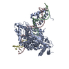
| ||||||||
|---|---|---|---|---|---|---|---|---|---|
| 1 | 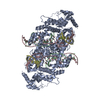
| ||||||||
| Unit cell |
|
-Components
| #1: Protein | Mass: 59033.633 Da / Num. of mol.: 1 / Mutation: C519S Source method: isolated from a genetically manipulated source Source: (gene. exp.) Musca domestica (house fly) / Production host:  |
|---|---|
| #2: DNA chain | Mass: 4942.273 Da / Num. of mol.: 1 / Source method: obtained synthetically / Source: (synth.) Musca domestica (house fly) |
| #3: DNA chain | Mass: 7983.107 Da / Num. of mol.: 1 / Source method: obtained synthetically / Source: (synth.) Musca domestica (house fly) |
| #4: DNA chain | Mass: 2427.605 Da / Num. of mol.: 1 / Source method: obtained synthetically / Source: (synth.) Musca domestica (house fly) |
| #5: Chemical | ChemComp-CA /   Mass: 40.078 Da / Num. of mol.: 1 / Source method: obtained synthetically / Formula: Ca Mass: 40.078 Da / Num. of mol.: 1 / Source method: obtained synthetically / Formula: Ca |
-Experimental details
-Experiment
| Experiment | Method: X-RAY DIFFRACTION / Number of used crystals: 1 |
|---|
- Sample preparation
| Crystal | Density Matthews: 2.85 Å3/Da / Density % sol: 56.79 % |
|---|---|
| Crystal grow | Temperature: 277 K / Method: vapor diffusion, hanging drop Details: 50 mM MES pH 5.8-6.5, 50 mM sodium acetate, 15% PEG MME 2000 |
-Data collection
| Diffraction | Mean temperature: 95 K |
|---|---|
| Diffraction source | Source: SYNCHROTRON / Site: APS  / Beamline: 22-ID / Wavelength: 1.5498 Å / Beamline: 22-ID / Wavelength: 1.5498 Å |
| Detector | Type: RAYONIX MX-300 / Detector: CCD / Date: Apr 13, 2013 |
| Radiation | Protocol: SINGLE WAVELENGTH / Monochromatic (M) / Laue (L): M / Scattering type: x-ray |
| Radiation wavelength | Wavelength: 1.5498 Å / Relative weight: 1 |
| Reflection | Resolution: 3.1→30 Å / Num. obs: 15708 / % possible obs: 99.5 % / Redundancy: 7.24 % / Rmerge(I) obs: 0.108 / Net I/σ(I): 13.61 |
| Reflection shell | Resolution: 3.1→3.18 Å / Rmerge(I) obs: 1.675 / Num. unique obs: 1152 |
- Processing
| Software |
| |||||||||||||||||||||||||||||||||||||||||||||||||
|---|---|---|---|---|---|---|---|---|---|---|---|---|---|---|---|---|---|---|---|---|---|---|---|---|---|---|---|---|---|---|---|---|---|---|---|---|---|---|---|---|---|---|---|---|---|---|---|---|---|---|
| Refinement | Method to determine structure: MOLECULAR REPLACEMENT Starting model: 4D1Q Resolution: 3.2→29.554 Å / SU ML: 0.43 / Cross valid method: FREE R-VALUE / σ(F): 1.99 / Phase error: 37.07
| |||||||||||||||||||||||||||||||||||||||||||||||||
| Solvent computation | Shrinkage radii: 1.1 Å / VDW probe radii: 1.3 Å | |||||||||||||||||||||||||||||||||||||||||||||||||
| Refinement step | Cycle: LAST / Resolution: 3.2→29.554 Å
| |||||||||||||||||||||||||||||||||||||||||||||||||
| Refine LS restraints |
| |||||||||||||||||||||||||||||||||||||||||||||||||
| LS refinement shell |
|
