 Movie
Movie Controller


+ Open data

- Basic information
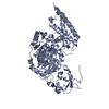
| Entry | Database: PDB / ID: 6ib8 | ||||||
|---|---|---|---|---|---|---|---|
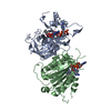
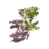
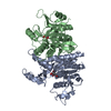
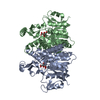
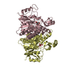
| Title | Structure of a complex of SuhB and NusA AR2 domain | ||||||
 Components Components |
| ||||||
 Keywords Keywords | TRANSCRIPTION / SuhB / NusA AR2 domain / antitermiantion | ||||||
| Function / homology |  Function and homology information Function and homology informationglycerol-2-phosphatase activity / inositol monophosphate 3-phosphatase activity / lithium ion binding / inositol-phosphate phosphatase / inositol monophosphate 1-phosphatase activity / inositol metabolic process / rRNA primary transcript binding / phosphatidylinositol phosphate biosynthetic process / protein complex oligomerization / transcription antitermination factor activity, RNA binding ...glycerol-2-phosphatase activity / inositol monophosphate 3-phosphatase activity / lithium ion binding / inositol-phosphate phosphatase / inositol monophosphate 1-phosphatase activity / inositol metabolic process / rRNA primary transcript binding / phosphatidylinositol phosphate biosynthetic process / protein complex oligomerization / transcription antitermination factor activity, RNA binding / bacterial-type RNA polymerase core enzyme binding / regulation of DNA-templated transcription elongation / transcription elongation factor complex / transcription antitermination / DNA-templated transcription termination / ribosome biogenesis / DNA-binding transcription factor activity / protein domain specific binding / nucleotide binding / magnesium ion binding / signal transduction / RNA binding / cytoplasm / cytosol Similarity search - Function | ||||||
| Biological species |  | ||||||
| Method |  X-RAY DIFFRACTION / SYNCHROTRON / MOLECULAR REPLACEMENT / Resolution: 1.646 Å X-RAY DIFFRACTION / SYNCHROTRON / MOLECULAR REPLACEMENT / Resolution: 1.646 Å | ||||||
 Authors Authors | Huang, Y.H. / Loll, B. / Wahl, M.C. | ||||||
 Citation Citation | Journal: Nucleic Acids Res. / Year: 2019 Title: Structural basis for the function of SuhB as a transcription factor in ribosomal RNA synthesis. Authors: Huang, Y.H. / Said, N. / Loll, B. / Wahl, M.C. | ||||||
| History |
|
- Structure visualization
| Structure viewer | Molecule: MolmilJmol/JSmol |
|---|
- Downloads & links
-Download
| PDBx/mmCIF format | 6ib8.cif.gz | 140.4 KB | Display | PDBx/mmCIF format |
|---|---|---|---|---|
| PDB format | pdb6ib8.ent.gz | 107.9 KB | Display | PDB format |
| PDBx/mmJSON format | 6ib8.json.gz | Tree view | PDBx/mmJSON format | |
| Others |  Other downloads Other downloads |
-Validation report
| Arichive directory | https://data.pdbj.org/pub/pdb/validation_reports/ib/6ib8ftp://data.pdbj.org/pub/pdb/validation_reports/ib/6ib8 | HTTPS FTP |
|---|
-Related structure data
| Related structure data |  6ib7C  1wcnS  2qflS S: Starting model for refinement C: citing same article ( |
|---|---|
| Similar structure data |


-Links
 PDBj
PDBj


- Assembly
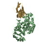
| Deposited unit | 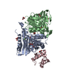
| ||||||||
|---|---|---|---|---|---|---|---|---|---|
| 1 |
| ||||||||
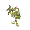
| Unit cell |
|
-Components
-Protein , 2 types, 3 molecules ABC
| #1: Protein | Mass: 29537.502 Da / Num. of mol.: 2 Source method: isolated from a genetically manipulated source Source: (gene. exp.) #2: Protein | | Mass: 7671.543 Da / Num. of mol.: 1 Source method: isolated from a genetically manipulated source Source: (gene. exp.) |
|---|
-Non-polymers , 4 types, 275 molecules 


| #3: Chemical | ChemComp-GOL /  Mass: 92.094 Da / Num. of mol.: 8 / Source method: obtained synthetically / Formula: C3H8O3 Mass: 92.094 Da / Num. of mol.: 8 / Source method: obtained synthetically / Formula: C3H8O3#4: Chemical |  Mass: 24.305 Da / Num. of mol.: 2 / Source method: obtained synthetically / Formula: Mg Mass: 24.305 Da / Num. of mol.: 2 / Source method: obtained synthetically / Formula: Mg#5: Chemical |  Mass: 354.436 Da / Num. of mol.: 2 / Source method: obtained synthetically / Formula: C16H34O8 / Comment: precipitant*YM Mass: 354.436 Da / Num. of mol.: 2 / Source method: obtained synthetically / Formula: C16H34O8 / Comment: precipitant*YM#6: Water | ChemComp-HOH / | Mass: 18.015 Da / Num. of mol.: 263 / Source method: isolated from a natural source / Formula: H2O |
|---|
-Experimental details
-Experiment
| Experiment | Method: X-RAY DIFFRACTION / Number of used crystals: 1 |
|---|
- Sample preparation
| Crystal | Density Matthews: 2.4 Å3/Da / Density % sol: 48.84 % |
|---|---|
| Crystal grow | Temperature: 291 K / Method: vapor diffusion, sitting drop / pH: 8.5 Details: 0.1M Tris-HCl pH 8.5, 5% (w/v) PEG 8000, 16% (v/v) PEG 300, 10% (v/v) glycerol |
-Data collection
| Diffraction | Mean temperature: 100 K / Serial crystal experiment: N |
|---|---|
| Diffraction source | Source: SYNCHROTRON / Site: BESSY  / Beamline: 14.2 / Wavelength: 0.91841 Å / Beamline: 14.2 / Wavelength: 0.91841 Å |
| Detector | Type: DECTRIS PILATUS 2M / Detector: PIXEL / Date: Oct 6, 2017 |
| Radiation | Monochromator: KMC-2 / Protocol: SINGLE WAVELENGTH / Monochromatic (M) / Laue (L): M / Scattering type: x-ray |
| Radiation wavelength | Wavelength: 0.91841 Å / Relative weight: 1 |
| Reflection | Resolution: 1.646→50 Å / Num. obs: 78162 / % possible obs: 99.1 % / Redundancy: 5.5 % / CC1/2: 0.99 / Net I/σ(I): 15.2 |
| Reflection shell | Resolution: 1.646→1.74 Å / Redundancy: 5.1 % / Mean I/σ(I) obs: 0.92 / Num. unique obs: 12069 / CC1/2: 0.36 / Rrim(I) all: 1.86 / % possible all: 95.7 |
- Processing
| Software |
| ||||||||||||||||||||||||||||||||||||||||||||||||||||||||||||||||||||||||||||||||||||||||||||||||||||||||||||||||
|---|---|---|---|---|---|---|---|---|---|---|---|---|---|---|---|---|---|---|---|---|---|---|---|---|---|---|---|---|---|---|---|---|---|---|---|---|---|---|---|---|---|---|---|---|---|---|---|---|---|---|---|---|---|---|---|---|---|---|---|---|---|---|---|---|---|---|---|---|---|---|---|---|---|---|---|---|---|---|---|---|---|---|---|---|---|---|---|---|---|---|---|---|---|---|---|---|---|---|---|---|---|---|---|---|---|---|---|---|---|---|---|---|---|
| Refinement | Method to determine structure: MOLECULAR REPLACEMENT Starting model: 2QFL, 1WCN Resolution: 1.646→30.634 Å / SU ML: 0.25 / Cross valid method: FREE R-VALUE / σ(F): 1.34 / Phase error: 22.84
| ||||||||||||||||||||||||||||||||||||||||||||||||||||||||||||||||||||||||||||||||||||||||||||||||||||||||||||||||
| Solvent computation | Shrinkage radii: 0.9 Å / VDW probe radii: 1.11 Å | ||||||||||||||||||||||||||||||||||||||||||||||||||||||||||||||||||||||||||||||||||||||||||||||||||||||||||||||||
| Refinement step | Cycle: LAST / Resolution: 1.646→30.634 Å
| ||||||||||||||||||||||||||||||||||||||||||||||||||||||||||||||||||||||||||||||||||||||||||||||||||||||||||||||||
| Refine LS restraints |
| ||||||||||||||||||||||||||||||||||||||||||||||||||||||||||||||||||||||||||||||||||||||||||||||||||||||||||||||||
| LS refinement shell |
|
