 Movie
Movie Controller

[English] 日本語
 Yorodumi
- PDB-6ht6: Crystal structure of glutathione transferase Omega 2S from Tramet... -
+ Open data

- Basic information
| Entry | Database: PDB / ID: 6ht6 | ||||||
|---|---|---|---|---|---|---|---|
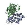
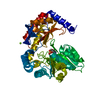
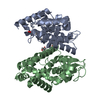
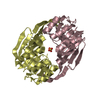
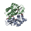
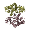
| Title | Crystal structure of glutathione transferase Omega 2S from Trametes versicolor in complex with 2,4-dihydroxybenzophenone | ||||||
 Components Components | Glutathione S-transferase omega 2S | ||||||
 Keywords Keywords | TRANSFERASE / glutathione transferase | ||||||
| Function / homology |  Function and homology information Function and homology information | ||||||
| Biological species |  Trametes versicolor (fungus) Trametes versicolor (fungus) | ||||||
| Method |  X-RAY DIFFRACTION / SYNCHROTRON / MOLECULAR REPLACEMENT / Resolution: 2.671 Å X-RAY DIFFRACTION / SYNCHROTRON / MOLECULAR REPLACEMENT / Resolution: 2.671 Å | ||||||
 Authors Authors | Schwartz, M. / Favier, F. / Didierjean, C. | ||||||
| Funding support |  France, 1items France, 1items
| ||||||
 Citation Citation | Journal: Sci Rep / Year: 2018 Title: Molecular recognition of wood polyphenols by phase II detoxification enzymes of the white rot Trametes versicolor. Authors: Schwartz, M. / Perrot, T. / Aubert, E. / Dumarcay, S. / Favier, F. / Gerardin, P. / Morel-Rouhier, M. / Mulliert, G. / Saiag, F. / Didierjean, C. / Gelhaye, E. | ||||||
| History |
|
- Structure visualization
| Structure viewer | Molecule: MolmilJmol/JSmol |
|---|
- Downloads & links
-Download
| PDBx/mmCIF format | 6ht6.cif.gz | 177.8 KB | Display | PDBx/mmCIF format |
|---|---|---|---|---|
| PDB format | pdb6ht6.ent.gz | 142.9 KB | Display | PDB format |
| PDBx/mmJSON format | 6ht6.json.gz | Tree view | PDBx/mmJSON format | |
| Others |  Other downloads Other downloads |
-Validation report
| Summary document | 6ht6_validation.pdf.gz | 458 KB | Display | wwPDB validaton report |
|---|---|---|---|---|
| Full document | 6ht6_full_validation.pdf.gz | 462.6 KB | Display | |
| Data in XML | 6ht6_validation.xml.gz | 17.9 KB | Display | |
| Data in CIF | 6ht6_validation.cif.gz | 24 KB | Display | |
| Arichive directory | https://data.pdbj.org/pub/pdb/validation_reports/ht/6ht6ftp://data.pdbj.org/pub/pdb/validation_reports/ht/6ht6 | HTTPS FTP |
-Related structure data
| Related structure data |  6f43C 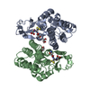 6f4bC 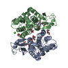 6f4fC  6f4kC  6f51C  6f66C 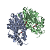 6f67C 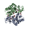 6f68C  6f69C  6f6aC  6f70C  6f71C 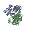 6gibS S: Starting model for refinement C: citing same article ( |
|---|---|
| Similar structure data |


-Links
 PDBj
PDBj

- Assembly
| Deposited unit | 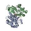
| ||||||||
|---|---|---|---|---|---|---|---|---|---|
| 1 |
| ||||||||
| Unit cell |
|
-Components
| #1: Protein | Mass: 27328.105 Da / Num. of mol.: 2 Source method: isolated from a genetically manipulated source Source: (gene. exp.) Trametes versicolor (fungus) / Production host:  #2: Chemical |   Mass: 214.217 Da / Num. of mol.: 2 / Source method: obtained synthetically / Formula: C13H10O3 Mass: 214.217 Da / Num. of mol.: 2 / Source method: obtained synthetically / Formula: C13H10O3#3: Chemical | ChemComp-MG / |   Mass: 24.305 Da / Num. of mol.: 1 / Source method: obtained synthetically / Formula: Mg Mass: 24.305 Da / Num. of mol.: 1 / Source method: obtained synthetically / Formula: Mg#4: Water | ChemComp-HOH / |  Mass: 18.015 Da / Num. of mol.: 16 / Source method: isolated from a natural source / Formula: H2O Mass: 18.015 Da / Num. of mol.: 16 / Source method: isolated from a natural source / Formula: H2O |
|---|
-Experimental details
-Experiment
| Experiment | Method: X-RAY DIFFRACTION / Number of used crystals: 1 |
|---|
- Sample preparation
| Crystal | Density Matthews: 2.4 Å3/Da / Density % sol: 48.84 % |
|---|---|
| Crystal grow | Temperature: 277 K / Method: vapor diffusion, hanging drop Details: 10.7% PEG4000, 0.1 M pH 7.0 HEPES-MES buffer (in the ratio 4:6, respectively), 0.05 M sodium acetate and 0.05 M magnesium chloride |
-Data collection
| Diffraction | Mean temperature: 100 K / Serial crystal experiment: N |
|---|---|
| Diffraction source | Source: SYNCHROTRON / Site: ESRF / Beamline: BM30A / Wavelength: 0.97951 Å |
| Detector | Type: ADSC QUANTUM 315r / Detector: CCD / Date: Dec 21, 2017 |
| Radiation | Protocol: SINGLE WAVELENGTH / Monochromatic (M) / Laue (L): M / Scattering type: x-ray |
| Radiation wavelength | Wavelength: 0.97951 Å / Relative weight: 1 |
| Reflection | Resolution: 2.67→47.774 Å / Num. obs: 15526 / % possible obs: 99.9 % / Redundancy: 7.2 % / Rmerge(I) obs: 0.137 / Rpim(I) all: 0.055 / Rrim(I) all: 0.148 / Net I/σ(I): 11.9 |
| Reflection shell | Resolution: 2.67→2.8 Å / Rmerge(I) obs: 0.619 / Mean I/σ(I) obs: 3.2 / Rpim(I) all: 0.245 / Rrim(I) all: 0.666 / % possible all: 99.1 |
- Processing
| Software |
| |||||||||||||||||||||||||||||||||||||||||||||||||
|---|---|---|---|---|---|---|---|---|---|---|---|---|---|---|---|---|---|---|---|---|---|---|---|---|---|---|---|---|---|---|---|---|---|---|---|---|---|---|---|---|---|---|---|---|---|---|---|---|---|---|
| Refinement | Method to determine structure: MOLECULAR REPLACEMENT Starting model: 6GIB Resolution: 2.671→47.774 Å / SU ML: 0.31 / Cross valid method: FREE R-VALUE / σ(F): 1.34 / Phase error: 29.23 / Stereochemistry target values: ML
| |||||||||||||||||||||||||||||||||||||||||||||||||
| Solvent computation | Shrinkage radii: 0.9 Å / VDW probe radii: 1.11 Å / Solvent model: FLAT BULK SOLVENT MODEL | |||||||||||||||||||||||||||||||||||||||||||||||||
| Refinement step | Cycle: LAST / Resolution: 2.671→47.774 Å
| |||||||||||||||||||||||||||||||||||||||||||||||||
| Refine LS restraints |
| |||||||||||||||||||||||||||||||||||||||||||||||||
| LS refinement shell |
|
