 Movie
Movie Controller

[English] 日本語
 Yorodumi
- PDB-6h12: Crystal structure of TcACHE complexed to 1-(6-Oxo-1,2,3,4,6,10b-h... -
+ Open data

- Basic information
| Entry | Database: PDB / ID: 6h12 | ||||||
|---|---|---|---|---|---|---|---|
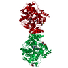
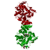
| Title | Crystal structure of TcACHE complexed to 1-(6-Oxo-1,2,3,4,6,10b-hexahydropyrido[2,1-a]isoindol-10-yl)-3-(4-(((1-(2-((1,2,3,4-tetrahydroacridin-9-yl)amino)ethyl)-1H-1,2,3-triazol-4-yl)methoxy)methyl)pyridin-2-yl)urea | ||||||
 Components Components | Acetylcholinesterase | ||||||
 Keywords Keywords | HYDROLASE / complex / inhibitor / alzheimer / acetylcholinesterase / multi-target-directed ligands / MTDL | ||||||
| Function / homology |  Function and homology information Function and homology informationacetylcholine catabolic process in synaptic cleft / acetylcholinesterase / acetylcholinesterase activity / synaptic cleft / side of membrane / synapse / plasma membraneSimilarity search - Function | ||||||
| Biological species |   Tetronarce californica (Pacific electric ray) Tetronarce californica (Pacific electric ray) | ||||||
| Method | X-RAY DIFFRACTION / SYNCHROTRON / MOLECULAR REPLACEMENT / Resolution: 2.2 Å | ||||||
 Authors Authors | Coquelle, N. / Colletier, J.P. | ||||||
| Funding support |  France, 1items France, 1items
| ||||||
 Citation Citation | Journal: Eur.J.Med.Chem. / Year: 2019 Title: Design, biological evaluation and X-ray crystallography of nanomolar multifunctional ligands targeting simultaneously acetylcholinesterase and glycogen synthase kinase-3. Authors: Oukoloff, K. / Coquelle, N. / Bartolini, M. / Naldi, M. / Le Guevel, R. / Bach, S. / Josselin, B. / Ruchaud, S. / Catto, M. / Pisani, L. / Denora, N. / Iacobazzi, R.M. / Silman, I. / ...Authors: Oukoloff, K. / Coquelle, N. / Bartolini, M. / Naldi, M. / Le Guevel, R. / Bach, S. / Josselin, B. / Ruchaud, S. / Catto, M. / Pisani, L. / Denora, N. / Iacobazzi, R.M. / Silman, I. / Sussman, J.L. / Buron, F. / Colletier, J.P. / Jean, L. / Routier, S. / Renard, P.Y. | ||||||
| History |
|
- Structure visualization
| Structure viewer | Molecule: MolmilJmol/JSmol |
|---|
- Downloads & links
-Download
| PDBx/mmCIF format | 6h12.cif.gz | 254.4 KB | Display | PDBx/mmCIF format |
|---|---|---|---|---|
| PDB format | pdb6h12.ent.gz | 202.9 KB | Display | PDB format |
| PDBx/mmJSON format | 6h12.json.gz | Tree view | PDBx/mmJSON format | |
| Others |  Other downloads Other downloads |
-Validation report
| Arichive directory | https://data.pdbj.org/pub/pdb/validation_reports/h1/6h12ftp://data.pdbj.org/pub/pdb/validation_reports/h1/6h12 | HTTPS FTP |
|---|
-Related structure data
| Related structure data |  6h13C  6h14C  2xi4S S: Starting model for refinement C: citing same article ( |
|---|---|
| Similar structure data |


-Links
 PDBj
PDBj
- Assembly
| Deposited unit | 
| ||||||||
|---|---|---|---|---|---|---|---|---|---|
| 1 | 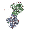
| ||||||||
| Unit cell |
|
-Components
-Protein / Sugars , 2 types, 7 molecules AB
| #1: Protein | / AChE Mass: 63763.965 Da / Num. of mol.: 2 / Source method: isolated from a natural source Source: (natural) Tetronarce californica (Pacific electric ray)References: UniProt: P04058, acetylcholinesterase#8: Sugar | ChemComp-NAG / N-Acetylglucosamine Type: D-saccharide, beta linking / Mass: 221.208 Da / Num. of mol.: 5 Type: D-saccharide, beta linking / Mass: 221.208 Da / Num. of mol.: 5Source method: isolated from a genetically manipulated source Formula: C8H15NO6 |
|---|
-Non-polymers , 9 types, 787 molecules 


| #2: Chemical | ChemComp-MES / MES (buffer) Mass: 195.237 Da / Num. of mol.: 4 Mass: 195.237 Da / Num. of mol.: 4Source method: isolated from a genetically manipulated source Formula: C6H13NO4S / Comment: pH buffer*YM #3: Chemical | Glycerol Mass: 92.094 Da / Num. of mol.: 3 / Source method: obtained synthetically / Formula: C3H8O3 Mass: 92.094 Da / Num. of mol.: 3 / Source method: obtained synthetically / Formula: C3H8O3#4: Chemical | Polyethylene glycol Mass: 150.173 Da / Num. of mol.: 2 / Source method: obtained synthetically / Formula: C6H14O4 Mass: 150.173 Da / Num. of mol.: 2 / Source method: obtained synthetically / Formula: C6H14O4#5: Chemical | ChemComp-EDO / Ethylene glycol Mass: 62.068 Da / Num. of mol.: 13 / Source method: obtained synthetically / Formula: C2H6O2 Mass: 62.068 Da / Num. of mol.: 13 / Source method: obtained synthetically / Formula: C2H6O2#6: Chemical | Sulfate Mass: 96.063 Da / Num. of mol.: 3 / Source method: isolated from a natural source / Formula: SO4 Mass: 96.063 Da / Num. of mol.: 3 / Source method: isolated from a natural source / Formula: SO4#7: Chemical | ChemComp-PEG / Diethylene glycol Mass: 106.120 Da / Num. of mol.: 8 / Source method: obtained synthetically / Formula: C4H10O3 Mass: 106.120 Da / Num. of mol.: 8 / Source method: obtained synthetically / Formula: C4H10O3#9: Chemical | ChemComp-CL / Chloride Mass: 35.453 Da / Num. of mol.: 6 / Source method: obtained synthetically / Formula: Cl Mass: 35.453 Da / Num. of mol.: 6 / Source method: obtained synthetically / Formula: Cl#10: Chemical |  Mass: 659.780 Da / Num. of mol.: 2 / Source method: obtained synthetically / Formula: C37H41N9O3 Mass: 659.780 Da / Num. of mol.: 2 / Source method: obtained synthetically / Formula: C37H41N9O3#11: Water | ChemComp-HOH / | WaterMass: 18.015 Da / Num. of mol.: 746 / Source method: isolated from a natural source / Formula: H2O |
|---|
-Experimental details
-Experiment
| Experiment | Method: X-RAY DIFFRACTION / Number of used crystals: 1 |
|---|
- Sample preparation
| Crystal | Density Matthews: 2.72 Å3/Da / Density % sol: 54.79 % |
|---|---|
| Crystal grow | Temperature: 277 K / Method: vapor diffusion, hanging drop / pH: 6 / Details: 30% PEG 200/50 mM MES pH 6.0 |
-Data collection
| Diffraction | Mean temperature: 100 K / Serial crystal experiment: N |
|---|---|
| Diffraction source | Source: SYNCHROTRON / Site: ESRF / Beamline: ID29 / Wavelength: 0.8726 Å |
| Detector | Type: MARMOSAIC 225 mm CCD / Detector: CCD / Date: Dec 8, 2013 |
| Radiation | Protocol: SINGLE WAVELENGTH / Monochromatic (M) / Laue (L): M / Scattering type: x-ray |
| Radiation wavelength | Wavelength: 0.8726 Å / Relative weight: 1 |
| Reflection | Resolution: 2.2→19.84 Å / Num. obs: 73534 / % possible obs: 99.63 % / Redundancy: 5.7 % / CC1/2: 0.999 / Rmerge(I) obs: 0.092 / Net I/σ(I): 13.6 |
| Reflection shell | Resolution: 2.2→2.26 Å / Redundancy: 6.3 % / Rmerge(I) obs: 0.587 / Mean I/σ(I) obs: 2.8 / Num. unique obs: 3584 / CC1/2: 0.995 / % possible all: 99.78 |
- Processing
| Software |
| |||||||||||||||||||||||||||||||||||||||||||||||||||||||||||||||||||||||||||||||||||||||||||||||||||||||||
|---|---|---|---|---|---|---|---|---|---|---|---|---|---|---|---|---|---|---|---|---|---|---|---|---|---|---|---|---|---|---|---|---|---|---|---|---|---|---|---|---|---|---|---|---|---|---|---|---|---|---|---|---|---|---|---|---|---|---|---|---|---|---|---|---|---|---|---|---|---|---|---|---|---|---|---|---|---|---|---|---|---|---|---|---|---|---|---|---|---|---|---|---|---|---|---|---|---|---|---|---|---|---|---|---|---|---|
| Refinement | Method to determine structure: MOLECULAR REPLACEMENT Starting model: 2xi4 Resolution: 2.2→19.84 Å / SU ML: 0.2 / Cross valid method: FREE R-VALUE / σ(F): 1.36 / Phase error: 22.97
| |||||||||||||||||||||||||||||||||||||||||||||||||||||||||||||||||||||||||||||||||||||||||||||||||||||||||
| Solvent computation | Shrinkage radii: 0.9 Å / VDW probe radii: 1.11 Å | |||||||||||||||||||||||||||||||||||||||||||||||||||||||||||||||||||||||||||||||||||||||||||||||||||||||||
| Refinement step | Cycle: LAST / Resolution: 2.2→19.84 Å
| |||||||||||||||||||||||||||||||||||||||||||||||||||||||||||||||||||||||||||||||||||||||||||||||||||||||||
| Refine LS restraints |
| |||||||||||||||||||||||||||||||||||||||||||||||||||||||||||||||||||||||||||||||||||||||||||||||||||||||||
| LS refinement shell |
|
