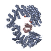
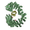
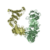
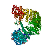
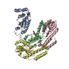
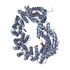
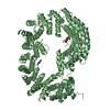
[English] 日本語
Yorodumi
- PDB-6gx9: Crystal structure of the TNPO3 - CPSF6 RSLD complex -

+
Open data


ID or keywords:

Loading...

-
Basic information

Entry
Database: PDB / ID: 6gx9
TitleCrystal structure of the TNPO3 - CPSF6 RSLD complex
Components
  • Cleavage and polyadenylation specificity factor subunit 6
  • Transportin-3
KeywordsNUCLEAR PROTEIN / karyopherin-cargo complex / nuclear import / SR-like domain
Function / homology
Function and homology information


exon-exon junction complex binding / : / positive regulation of RNA export from nucleus / mRNA cleavage factor complex / annulate lamellae / interchromatin granule / Processing of Intronless Pre-mRNAs / perichromatin fibrils / mRNA cleavage and polyadenylation specificity factor complex / mRNA alternative polyadenylation ...exon-exon junction complex binding / : / positive regulation of RNA export from nucleus / mRNA cleavage factor complex / annulate lamellae / interchromatin granule / Processing of Intronless Pre-mRNAs / perichromatin fibrils / mRNA cleavage and polyadenylation specificity factor complex / mRNA alternative polyadenylation / paraspeckles / mRNA 3'-end processing / mRNA 3'-end processing / Signaling by cytosolic FGFR1 fusion mutants / RNA Polymerase II Transcription Termination / protein heterotetramerization / nuclear import signal receptor activity / ribosomal large subunit binding / Processing of Capped Intron-Containing Pre-mRNA / localization / Signaling by FGFR1 in disease / protein tetramerization / mRNA processing / small GTPase binding / protein import into nucleus / nuclear envelope / nuclear speck / ribonucleoprotein complex / mRNA binding / intracellular membrane-bounded organelle / RNA binding / nucleoplasm / membrane / identical protein binding / nucleus / cytoplasm
Similarity search - Function
Cleavage and polyadenylation specificity factor subunit 6 / CPSF6/7 family / Exportin-1/Importin-beta-like / Exportin 1-like protein / RNA recognition motif / RNA recognition motif / Eukaryotic RNA Recognition Motif (RRM) profile. / RNA recognition motif domain / RNA-binding domain superfamily / Armadillo-like helical ...Cleavage and polyadenylation specificity factor subunit 6 / CPSF6/7 family / Exportin-1/Importin-beta-like / Exportin 1-like protein / RNA recognition motif / RNA recognition motif / Eukaryotic RNA Recognition Motif (RRM) profile. / RNA recognition motif domain / RNA-binding domain superfamily / Armadillo-like helical / Armadillo-type fold / Nucleotide-binding alpha-beta plait domain superfamily
Similarity search - Domain/homology
BENZAMIDINE / Cleavage and polyadenylation specificity factor subunit 6 / Transportin-3
Similarity search - Component
Biological speciesHomo sapiens (human)
MethodX-RAY DIFFRACTION / SYNCHROTRON / MOLECULAR REPLACEMENT / Resolution: 2.7 Å
AuthorsCherepanov, P. / Cook, N.
Funding support United States, 1items
OrganizationGrant numberCountry
National Institutes of Health/National Institute of Diabetes and Digestive and Kidney Disease (NIH/NIDDK)GM082251 United States
CitationJournal: Nucleic Acids Res. / Year: 2019
Title: Differential role for phosphorylation in alternative polyadenylation function versus nuclear import of SR-like protein CPSF6.
Authors: Jang, S. / Cook, N.J. / Pye, V.E. / Bedwell, G.J. / Dudek, A.M. / Singh, P.K. / Cherepanov, P. / Engelman, A.N.
History
DepositionJun 26, 2018Deposition site: PDBE / Processing site: PDBE
Revision 1.0Mar 13, 2019Provider: repository / Type: Initial release
Revision 1.1Apr 10, 2019Group: Data collection / Database references / Category: citation / citation_author / pdbx_database_proc
Item: _citation.country / _citation.journal_abbrev ..._citation.country / _citation.journal_abbrev / _citation.journal_id_ASTM / _citation.journal_id_CSD / _citation.journal_id_ISSN / _citation.pdbx_database_id_DOI / _citation.pdbx_database_id_PubMed / _citation.title / _citation.year / _citation_author.name
Revision 1.2May 22, 2019Group: Data collection / Database references
Category: citation / database_PDB_rev ...citation / database_PDB_rev / database_PDB_rev_record / pdbx_database_proc
Item: _citation.journal_volume / _citation.page_first / _citation.page_last
Revision 1.3Mar 30, 2022Group: Author supporting evidence / Database references / Derived calculations
Category: database_2 / pdbx_audit_support ...database_2 / pdbx_audit_support / pdbx_struct_conn_angle / struct_conn
Item: _database_2.pdbx_DOI / _database_2.pdbx_database_accession ..._database_2.pdbx_DOI / _database_2.pdbx_database_accession / _pdbx_audit_support.funding_organization / _pdbx_struct_conn_angle.ptnr1_auth_asym_id / _pdbx_struct_conn_angle.ptnr1_auth_seq_id / _pdbx_struct_conn_angle.ptnr1_label_asym_id / _pdbx_struct_conn_angle.ptnr1_label_atom_id / _pdbx_struct_conn_angle.ptnr1_label_seq_id / _pdbx_struct_conn_angle.ptnr2_auth_asym_id / _pdbx_struct_conn_angle.ptnr2_auth_seq_id / _pdbx_struct_conn_angle.ptnr2_label_asym_id / _pdbx_struct_conn_angle.ptnr2_symmetry / _pdbx_struct_conn_angle.ptnr3_auth_asym_id / _pdbx_struct_conn_angle.ptnr3_auth_seq_id / _pdbx_struct_conn_angle.ptnr3_label_asym_id / _pdbx_struct_conn_angle.ptnr3_label_atom_id / _pdbx_struct_conn_angle.ptnr3_label_seq_id / _pdbx_struct_conn_angle.value / _struct_conn.conn_type_id / _struct_conn.id / _struct_conn.pdbx_dist_value / _struct_conn.pdbx_leaving_atom_flag / _struct_conn.ptnr1_auth_asym_id / _struct_conn.ptnr1_auth_comp_id / _struct_conn.ptnr1_auth_seq_id / _struct_conn.ptnr1_label_asym_id / _struct_conn.ptnr1_label_atom_id / _struct_conn.ptnr1_label_comp_id / _struct_conn.ptnr1_label_seq_id / _struct_conn.ptnr2_auth_asym_id / _struct_conn.ptnr2_auth_comp_id / _struct_conn.ptnr2_auth_seq_id / _struct_conn.ptnr2_label_asym_id / _struct_conn.ptnr2_label_atom_id / _struct_conn.ptnr2_label_comp_id / _struct_conn.ptnr2_label_seq_id / _struct_conn.ptnr2_symmetry
Revision 1.4Jan 17, 2024Group: Data collection / Refinement description
Category: chem_comp_atom / chem_comp_bond / pdbx_initial_refinement_model

-
Structure visualization

Structure viewerMolecule:
MolmilJmol/JSmol

Downloads & links

-
Assembly

Deposited unit
A: Transportin-3
B: Transportin-3
C: Cleavage and polyadenylation specificity factor subunit 6
D: Cleavage and polyadenylation specificity factor subunit 6
hetero molecules


Theoretical massNumber of molelcules
Total (without water)227,92012
Polymers227,2564
Non-polymers6648
Water54030
1
A: Transportin-3
C: Cleavage and polyadenylation specificity factor subunit 6
hetero molecules


Theoretical massNumber of molelcules
Total (without water)113,9606
Polymers113,6282
Non-polymers3324
Water181
TypeNameSymmetry operationNumber
identity operation1_555x,y,z1
Buried area2220 Å2
ΔGint-3 kcal/mol
Surface area40540 Å2
MethodPISA
2
B: Transportin-3
D: Cleavage and polyadenylation specificity factor subunit 6
hetero molecules


Theoretical massNumber of molelcules
Total (without water)113,9606
Polymers113,6282
Non-polymers3324
Water181
TypeNameSymmetry operationNumber
identity operation1_555x,y,z1
Buried area2240 Å2
ΔGint-3 kcal/mol
Surface area40730 Å2
MethodPISA
Unit cell
Length a, b, c (Å)200.335, 200.335, 234.807
Angle α, β, γ (deg.)90.00, 90.00, 120.00
Int Tables number182
Space group name H-MP6322

-
Components

-
Protein , 2 types, 4 molecules ABCD

#1: Protein Transportin-3 / / Importin-12 / Imp12 / Transportin-SR / TRN-SR


Mass: 104313.211 Da / Num. of mol.: 2
Source method: isolated from a genetically manipulated source
Source: (gene. exp.) Homo sapiens (human) / Gene: TNPO3, IPO12 / Plasmid: pETSUMO-TNPO3 / Production host: Escherichia coli (E. coli) / References: UniProt: Q9Y5L0
#2: Protein Cleavage and polyadenylation specificity factor subunit 6 / / Cleavage and polyadenylation specificity factor 68 kDa subunit / CPSF 68 kDa subunit / Cleavage ...Cleavage and polyadenylation specificity factor 68 kDa subunit / CPSF 68 kDa subunit / Cleavage factor Im complex 68 kDa subunit / CFIm68 / Pre-mRNA cleavage factor Im 68 kDa subunit / Protein HPBRII-4/7


Mass: 9314.888 Da / Num. of mol.: 2
Source method: isolated from a genetically manipulated source
Source: (gene. exp.) Homo sapiens (human) / Gene: CPSF6, CFIM68 / Production host: Escherichia coli (E. coli) / References: UniProt: Q16630

-
Non-polymers , 4 types, 38 molecules

#3: Chemical
ChemComp-MG / MAGNESIUM ION


Mass: 24.305 Da / Num. of mol.: 4 / Source method: obtained synthetically / Formula: Mg
#4: Chemical ChemComp-BCN / BICINE / Bicine


Mass: 163.172 Da / Num. of mol.: 2 / Source method: obtained synthetically / Formula: C6H13NO4 / Comment: pH buffer*YM
#5: Chemical ChemComp-BEN / BENZAMIDINE / Benzamidine


Mass: 120.152 Da / Num. of mol.: 2 / Source method: obtained synthetically / Formula: C7H8N2
#6: Water ChemComp-HOH / water / Water


Mass: 18.015 Da / Num. of mol.: 30 / Source method: isolated from a natural source / Formula: H2O

-
Experimental details

-
Experiment

ExperimentMethod: X-RAY DIFFRACTION / Number of used crystals: 1

-
Sample preparation

CrystalDensity Matthews: 2.99 Å3/Da / Density % sol: 58.9 %
Crystal growTemperature: 291 K / Method: vapor diffusion, hanging drop / pH: 8
Details: 12.8% PEG MME 500, 6.4% PEG 20,000, 4% 1,6-hexandiol, 1.5% benzamidine, 75 mM NaCl, 40 mM MgCl2, 6 mM DTT, 80 mM Tris-bicine, pH 8.0

-
Data collection

DiffractionMean temperature: 100 K
Diffraction sourceSource: SYNCHROTRON / Site: Diamond / Beamline: I03 / Wavelength: 0.97625 Å
DetectorType: DECTRIS PILATUS 6M / Detector: PIXEL / Date: Jul 5, 2014
RadiationProtocol: SINGLE WAVELENGTH / Monochromatic (M) / Laue (L): M / Scattering type: x-ray
Radiation wavelengthWavelength: 0.97625 Å / Relative weight: 1
ReflectionResolution: 2.7→50.69 Å / Num. obs: 76497 / % possible obs: 100 % / Redundancy: 12 % / Biso Wilson estimate: 65.17 Å2 / CC1/2: 0.998 / Rmerge(I) obs: 0.171 / Rpim(I) all: 0.051 / Rrim(I) all: 0.179 / Net I/σ(I): 11.6
Reflection shellResolution: 2.7→2.85 Å / Redundancy: 12 % / Rmerge(I) obs: 1.931 / Mean I/σ(I) obs: 1.5 / Num. unique obs: 10987 / CC1/2: 0.422 / Rpim(I) all: 0.579 / Rrim(I) all: 2.018 / % possible all: 100

-
Processing

Software
NameVersionClassification
PHENIX(1.13_2998: ???)refinement
MOSFLMdata reduction
SCALAdata scaling
PHASERphasing
RefinementMethod to determine structure: MOLECULAR REPLACEMENT
Starting model: 4C0O
Resolution: 2.7→50.646 Å / SU ML: 0.4 / Cross valid method: FREE R-VALUE / σ(F): 1.33 / Phase error: 27.02
RfactorNum. reflection% reflection
Rfree0.248 3194 4.18 %
Rwork0.2137 --
obs0.2152 76443 99.98 %
Solvent computationShrinkage radii: 0.9 Å / VDW probe radii: 1.11 Å
Refinement stepCycle: LAST / Resolution: 2.7→50.646 Å
ProteinNucleic acidLigandSolventTotal
Num. atoms14593 0 44 30 14667
Refine LS restraints
Refine-IDTypeDev idealNumber
X-RAY DIFFRACTIONf_bond_d0.00214919
X-RAY DIFFRACTIONf_angle_d0.43220250
X-RAY DIFFRACTIONf_dihedral_angle_d11.7989038
X-RAY DIFFRACTIONf_chiral_restr0.0352364
X-RAY DIFFRACTIONf_plane_restr0.0032591
LS refinement shell
Resolution (Å)Rfactor RfreeNum. reflection RfreeRfactor RworkNum. reflection RworkRefine-ID% reflection obs (%)
2.7-2.74030.32641190.29463139X-RAY DIFFRACTION100
2.7403-2.78310.29771280.29343167X-RAY DIFFRACTION100
2.7831-2.82870.38231540.31163101X-RAY DIFFRACTION100
2.8287-2.87750.33961150.32223137X-RAY DIFFRACTION100
2.8775-2.92980.36821420.30533146X-RAY DIFFRACTION100
2.9298-2.98620.29111400.29093140X-RAY DIFFRACTION100
2.9862-3.04710.34671380.28363143X-RAY DIFFRACTION100
3.0471-3.11340.32891260.27673153X-RAY DIFFRACTION100
3.1134-3.18580.29161400.27163149X-RAY DIFFRACTION100
3.1858-3.26540.28381390.25943136X-RAY DIFFRACTION100
3.2654-3.35370.3061410.25853165X-RAY DIFFRACTION100
3.3537-3.45240.35591400.26353156X-RAY DIFFRACTION100
3.4524-3.56380.28281380.24423142X-RAY DIFFRACTION100
3.5638-3.69110.27121410.2343177X-RAY DIFFRACTION100
3.6911-3.83890.28251410.21993176X-RAY DIFFRACTION100
3.8389-4.01350.22761410.19773174X-RAY DIFFRACTION100
4.0135-4.2250.21431410.18813185X-RAY DIFFRACTION100
4.225-4.48960.22561410.18513192X-RAY DIFFRACTION100
4.4896-4.8360.22141410.17853210X-RAY DIFFRACTION100
4.836-5.32210.24841440.18773234X-RAY DIFFRACTION100
5.3221-6.09120.22051440.21063248X-RAY DIFFRACTION100
6.0912-7.66990.22941460.19863290X-RAY DIFFRACTION100
7.6699-50.65450.1641540.15073489X-RAY DIFFRACTION100

+
About Yorodumi

-
News

-
Feb 9, 2022. New format data for meta-information of EMDB entries

New format data for meta-information of EMDB entries

  • Version 3 of the EMDB header file is now the official format.
  • The previous official version 1.9 will be removed from the archive.

Related info.:EMDB header

External links:wwPDB to switch to version 3 of the EMDB data model

-
Aug 12, 2020. Covid-19 info

Covid-19 info

URL: https://pdbj.org/emnavi/covid19.php

New page: Covid-19 featured information page in EM Navigator.

Related info.:Covid-19 info / Mar 5, 2020. Novel coronavirus structure data

+
Mar 5, 2020. Novel coronavirus structure data

Novel coronavirus structure data

Related info.:Yorodumi Speices / Aug 12, 2020. Covid-19 info

External links:COVID-19 featured content - PDBj / Molecule of the Month (242):Coronavirus Proteases

+
Jan 31, 2019. EMDB accession codes are about to change! (news from PDBe EMDB page)

EMDB accession codes are about to change! (news from PDBe EMDB page)

  • The allocation of 4 digits for EMDB accession codes will soon come to an end. Whilst these codes will remain in use, new EMDB accession codes will include an additional digit and will expand incrementally as the available range of codes is exhausted. The current 4-digit format prefixed with “EMD-” (i.e. EMD-XXXX) will advance to a 5-digit format (i.e. EMD-XXXXX), and so on. It is currently estimated that the 4-digit codes will be depleted around Spring 2019, at which point the 5-digit format will come into force.
  • The EM Navigator/Yorodumi systems omit the EMD- prefix.

Related info.:Q: What is EMD? / ID/Accession-code notation in Yorodumi/EM Navigator

External links:EMDB Accession Codes are Changing Soon! / Contact to PDBj

+
Jul 12, 2017. Major update of PDB

Major update of PDB

  • wwPDB released updated PDB data conforming to the new PDBx/mmCIF dictionary.
  • This is a major update changing the version number from 4 to 5, and with Remediation, in which all the entries are updated.
  • In this update, many items about electron microscopy experimental information are reorganized (e.g. em_software).
  • Now, EM Navigator and Yorodumi are based on the updated data.

External links:wwPDB Remediation / Enriched Model Files Conforming to OneDep Data Standards Now Available in the PDB FTP Archive

-
Yorodumi

Thousand views of thousand structures

  • Yorodumi is a browser for structure data from EMDB, PDB, SASBDB, etc.
  • This page is also the successor to EM Navigator detail page, and also detail information page/front-end page for Omokage search.
  • The word "yorodu" (or yorozu) is an old Japanese word meaning "ten thousand". "mi" (miru) is to see.

Related info.:EMDB / PDB / SASBDB / Comparison of 3 databanks / Yorodumi Search / Aug 31, 2016. New EM Navigator & Yorodumi / Yorodumi Papers / Jmol/JSmol / Function and homology information / Changes in new EM Navigator and Yorodumi

Read more