: / positive regulation of retrograde transport, endosome to Golgi / regulation of lipid transport / positive regulation of neurotransmitter uptake / negative regulation of endoplasmic reticulum stress-induced neuron intrinsic apoptotic signaling pathway / negative regulation of spontaneous neurotransmitter secretion / negative regulation of intralumenal vesicle formation / regulation protein catabolic process at presynapse / cellular response to L-glutamine / : ...: / positive regulation of retrograde transport, endosome to Golgi / regulation of lipid transport / positive regulation of neurotransmitter uptake / negative regulation of endoplasmic reticulum stress-induced neuron intrinsic apoptotic signaling pathway / negative regulation of spontaneous neurotransmitter secretion / negative regulation of intralumenal vesicle formation / regulation protein catabolic process at presynapse / cellular response to L-glutamine / : / negative regulation of exosomal secretion / mitochondrion to lysosome vesicle-mediated transport / type 2 mitophagy / negative regulation of glucokinase activity / response to curcumin / negative regulation of mitochondrial fusion / cellular response to hydrogen sulfide / protein K29-linked ubiquitination / free ubiquitin chain polymerization / Lewy body / positive regulation of protein linear polyubiquitination / host-mediated suppression of viral genome replication / Parkin-FBXW7-Cul1 ubiquitin ligase complex / RBR-type E3 ubiquitin transferase / regulation of synaptic vesicle transport / positive regulation of mitochondrial fusion / negative regulation of actin filament bundle assembly / positive regulation of mitophagy / F-box domain binding / regulation of necroptotic process / mitochondrial fragmentation involved in apoptotic process / regulation of cellular response to oxidative stress / positive regulation of dendrite extension / regulation of dopamine metabolic process / negative regulation of excitatory postsynaptic potential / protein K6-linked ubiquitination / norepinephrine metabolic process / dopaminergic synapse / autophagy of mitochondrion / positive regulation of type 2 mitophagy / mitochondrion localization / protein localization to mitochondrion / positive regulation of tumor necrosis factor-mediated signaling pathway / cellular response to dopamine / positive regulation of proteasomal protein catabolic process / positive regulation of protein localization to membrane / mitochondrial fission / cellular response to toxic substance / protein K27-linked ubiquitination / negative regulation of intrinsic apoptotic signaling pathway by p53 class mediator / aggresome assembly / negative regulation of oxidative stress-induced neuron intrinsic apoptotic signaling pathway / cellular response to L-glutamate / regulation of mitochondrion organization / protein K11-linked ubiquitination / hypothalamus gonadotrophin-releasing hormone neuron development / ubiquitin conjugating enzyme binding / negative regulation of synaptic transmission, glutamatergic / regulation of canonical Wnt signaling pathway / female meiosis I / positive regulation of protein monoubiquitination / positive regulation of mitochondrial membrane potential / negative regulation of JNK cascade / aggresome / fat pad development / mitochondrion transport along microtubule / regulation of reactive oxygen species metabolic process / positive regulation of mitochondrial fission / response to corticosterone / female gonad development / seminiferous tubule development / response to muscle activity / negative regulation of release of cytochrome c from mitochondria / ubiquitin-specific protease binding / startle response / dopamine metabolic process / dopamine uptake involved in synaptic transmission / regulation of dopamine secretion / positive regulation of ATP biosynthetic process / male meiosis I / negative regulation of reactive oxygen species metabolic process / regulation of glucose metabolic process / cullin family protein binding / regulation of protein ubiquitination / positive regulation of intrinsic apoptotic signaling pathway by p53 class mediator / protein deubiquitination / cellular response to unfolded protein / protein K63-linked ubiquitination / regulation of synaptic vesicle endocytosis / protein monoubiquitination / ubiquitin ligase complex / negative regulation of mitochondrial fission / positive regulation of insulin secretion involved in cellular response to glucose stimulus / negative regulation of endoplasmic reticulum stress-induced intrinsic apoptotic signaling pathway / regulation of postsynaptic membrane neurotransmitter receptor levels / protein K48-linked ubiquitination / protein autoubiquitination / mitophagy / energy homeostasis / proteasomal protein catabolic process Similarity search - Function
In the structure databanks used in Yorodumi, some data are registered as the other names, "COVID-19 virus" and "2019-nCoV". Here are the details of the virus and the list of structure data.
Jan 31, 2019. EMDB accession codes are about to change! (news from PDBe EMDB page)
EMDB accession codes are about to change! (news from PDBe EMDB page)
The allocation of 4 digits for EMDB accession codes will soon come to an end. Whilst these codes will remain in use, new EMDB accession codes will include an additional digit and will expand incrementally as the available range of codes is exhausted. The current 4-digit format prefixed with “EMD-” (i.e. EMD-XXXX) will advance to a 5-digit format (i.e. EMD-XXXXX), and so on. It is currently estimated that the 4-digit codes will be depleted around Spring 2019, at which point the 5-digit format will come into force.
The EM Navigator/Yorodumi systems omit the EMD- prefix.
Related info.:Q: What is EMD? / ID/Accession-code notation in Yorodumi/EM Navigator
Yorodumi is a browser for structure data from EMDB, PDB, SASBDB, etc.
This page is also the successor to EM Navigator detail page, and also detail information page/front-end page for Omokage search.
The word "yorodu" (or yorozu) is an old Japanese word meaning "ten thousand". "mi" (miru) is to see.
Related info.:EMDB / PDB / SASBDB / Comparison of 3 databanks / Yorodumi Search / Aug 31, 2016. New EM Navigator & Yorodumi / Yorodumi Papers / Jmol/JSmol / Function and homology information / Changes in new EM Navigator and Yorodumi
 Movie
Movie Controller


 Open data
 Basic information
 Components
 Keywords
 Function and homology information
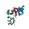
 Homo sapiens (human)
 X-RAY DIFFRACTION /
  Authors
 United Kingdom, 4items
  Citation
 Structure visualization
 Downloads & links
 Other downloads

 PDBj
PDBj


 Assembly
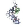


 Mass: 65.409 Da / Num. of mol.: 6 / Source method: obtained synthetically / Formula: Zn
 Mass: 92.094 Da / Num. of mol.: 3 / Source method: obtained synthetically / Formula: C3H8O3
 Mass: 118.174 Da / Num. of mol.: 1 / Source method: obtained synthetically / Formula: C6H14O2 / Comment: precipitant*YM
 Mass: 96.063 Da / Num. of mol.: 1 / Source method: obtained synthetically / Formula: SO4
 Sample preparation
 Processing