 Movie
Movie Controller

[English] 日本語
 Yorodumi
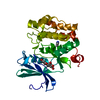
- PDB-6dgq: Crystal Structure of Human PPARgamma Ligand Binding Domain in Com... -
+ Open data

- Basic information
| Entry | Database: PDB / ID: 6dgq | ||||||
|---|---|---|---|---|---|---|---|
| Title | Crystal Structure of Human PPARgamma Ligand Binding Domain in Complex with CAY10506 | ||||||
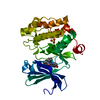
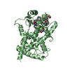
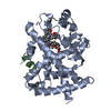
 Components Components | Peroxisome proliferator-activated receptor gamma | ||||||
 Keywords Keywords | TRANSCRIPTION/TRANSCRIPTION INHIBITOR / Nuclear receptors / TZDs / Drug design / Therapeutic targets / TRANSCRIPTION / TRANSCRIPTION-TRANSCRIPTION INHIBITOR complex | ||||||
| Function / homology |  Function and homology information Function and homology informationComplex I biogenesis / Respiratory electron transport / mitochondrial ATP synthesis coupled electron transport / mitochondrial respiratory chain complex I assembly / : / mitochondrial electron transport, NADH to ubiquinone / proton motive force-driven mitochondrial ATP synthesis / Mitochondrial protein degradation / NADH dehydrogenase (ubiquinone) activity / mitochondrial membrane ...Complex I biogenesis / Respiratory electron transport / mitochondrial ATP synthesis coupled electron transport / mitochondrial respiratory chain complex I assembly / : / mitochondrial electron transport, NADH to ubiquinone / proton motive force-driven mitochondrial ATP synthesis / Mitochondrial protein degradation / NADH dehydrogenase (ubiquinone) activity / mitochondrial membrane / aerobic respiration / mitochondrial inner membrane / mitochondrion / nucleoplasm Similarity search - Function | ||||||
| Biological species |  Homo sapiens (human) Homo sapiens (human) | ||||||
| Method |  X-RAY DIFFRACTION / SYNCHROTRON / MOLECULAR REPLACEMENT / Resolution: 2.45 Å X-RAY DIFFRACTION / SYNCHROTRON / MOLECULAR REPLACEMENT / Resolution: 2.45 Å | ||||||
 Authors Authors | Shang, J. / Kojetin, D.J. | ||||||
| Funding support |  United States, 1items United States, 1items
| ||||||
 Citation Citation | Journal: Proc.Natl.Acad.Sci.USA / Year: 2019 Title: Quantitative structural assessment of graded receptor agonism. Authors: Shang, J. / Brust, R. / Griffin, P.R. / Kamenecka, T.M. / Kojetin, D.J. | ||||||
| History |
|
- Structure visualization
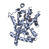
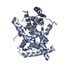
| Structure viewer | Molecule: MolmilJmol/JSmol |
|---|
- Downloads & links
-Download
| PDBx/mmCIF format | 6dgq.cif.gz | 118.1 KB | Display | PDBx/mmCIF format |
|---|---|---|---|---|
| PDB format | pdb6dgq.ent.gz | 90.4 KB | Display | PDB format |
| PDBx/mmJSON format | 6dgq.json.gz | Tree view | PDBx/mmJSON format | |
| Others |  Other downloads Other downloads |
-Validation report
| Summary document | 6dgq_validation.pdf.gz | 717.6 KB | Display | wwPDB validaton report |
|---|---|---|---|---|
| Full document | 6dgq_full_validation.pdf.gz | 731.1 KB | Display | |
| Data in XML | 6dgq_validation.xml.gz | 23.3 KB | Display | |
| Data in CIF | 6dgq_validation.cif.gz | 32.5 KB | Display | |
| Arichive directory | https://data.pdbj.org/pub/pdb/validation_reports/dg/6dgqftp://data.pdbj.org/pub/pdb/validation_reports/dg/6dgq | HTTPS FTP |
-Related structure data
| Related structure data |  6dglC  6dgoC  6dgrC  6o67C  6o68C 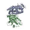 1prgS S: Starting model for refinement C: citing same article ( |
|---|---|
| Similar structure data |


-Links
 PDBj
PDBj- Assembly
| Deposited unit | 
| ||||||||
|---|---|---|---|---|---|---|---|---|---|
| 1 | 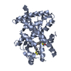
| ||||||||
| 2 | 
| ||||||||
| Unit cell |
|
-Components
| #1: Protein | Mass: 31506.574 Da / Num. of mol.: 2 Source method: isolated from a genetically manipulated source Source: (gene. exp.) Homo sapiens (human) / Gene: PPARG, NR1C3 / Plasmid: pET46 / Production host:  #2: Chemical | ChemComp-GBV / |   Mass: 454.626 Da / Num. of mol.: 1 / Source method: obtained synthetically / Formula: C20H26N2O4S3 Mass: 454.626 Da / Num. of mol.: 1 / Source method: obtained synthetically / Formula: C20H26N2O4S3#3: Water | ChemComp-HOH / |  Mass: 18.015 Da / Num. of mol.: 185 / Source method: isolated from a natural source / Formula: H2O Mass: 18.015 Da / Num. of mol.: 185 / Source method: isolated from a natural source / Formula: H2O |
|---|
-Experimental details
-Experiment
| Experiment | Method: X-RAY DIFFRACTION / Number of used crystals: 1 |
|---|
- Sample preparation
| Crystal | Density Matthews: 2.47 Å3/Da / Density % sol: 50.19 % |
|---|---|
| Crystal grow | Temperature: 293 K / Method: vapor diffusion, sitting drop / Details: 0.8M SODIUM CITRATE, 100mM Tris, pH 7.6 |
-Data collection
| Diffraction | Mean temperature: 100 K |
|---|---|
| Diffraction source | Source: SYNCHROTRON / Site: ALS / Beamline: 5.0.2 / Wavelength: 0.97741 Å |
| Detector | Type: DECTRIS PILATUS3 6M / Detector: PIXEL / Date: Aug 16, 2017 |
| Radiation | Monochromator: Double-crystal, Si(111) / Protocol: SINGLE WAVELENGTH / Monochromatic (M) / Laue (L): M / Scattering type: x-ray |
| Radiation wavelength | Wavelength: 0.97741 Å / Relative weight: 1 |
| Reflection | Resolution: 2.45→45.48 Å / Num. obs: 22240 / % possible obs: 97.3 % / Redundancy: 2 % / CC1/2: 0.998 / Net I/σ(I): 12.38 |
| Reflection shell | Resolution: 2.45→2.538 Å / Redundancy: 2 % / Mean I/σ(I) obs: 2.89 / Num. unique obs: 2193 / CC1/2: 0.811 / % possible all: 96.95 |
- Processing
| Software |
| |||||||||||||||||||||||||||||||||||||||||||||||||||||||||||||||||||||||||||||||||||||||||||||||||||||||||
|---|---|---|---|---|---|---|---|---|---|---|---|---|---|---|---|---|---|---|---|---|---|---|---|---|---|---|---|---|---|---|---|---|---|---|---|---|---|---|---|---|---|---|---|---|---|---|---|---|---|---|---|---|---|---|---|---|---|---|---|---|---|---|---|---|---|---|---|---|---|---|---|---|---|---|---|---|---|---|---|---|---|---|---|---|---|---|---|---|---|---|---|---|---|---|---|---|---|---|---|---|---|---|---|---|---|---|
| Refinement | Method to determine structure: MOLECULAR REPLACEMENT Starting model: 1PRG Resolution: 2.45→45.48 Å / SU ML: 0.31 / Cross valid method: FREE R-VALUE / σ(F): 1.37 / Phase error: 24.51
| |||||||||||||||||||||||||||||||||||||||||||||||||||||||||||||||||||||||||||||||||||||||||||||||||||||||||
| Solvent computation | Shrinkage radii: 0.9 Å / VDW probe radii: 1.11 Å | |||||||||||||||||||||||||||||||||||||||||||||||||||||||||||||||||||||||||||||||||||||||||||||||||||||||||
| Refinement step | Cycle: LAST / Resolution: 2.45→45.48 Å
| |||||||||||||||||||||||||||||||||||||||||||||||||||||||||||||||||||||||||||||||||||||||||||||||||||||||||
| Refine LS restraints |
| |||||||||||||||||||||||||||||||||||||||||||||||||||||||||||||||||||||||||||||||||||||||||||||||||||||||||
| LS refinement shell |
|
