 Movie
Movie Controller

[English] 日本語
 Yorodumi
- PDB-6d5c: Structure of Caldicellulosiruptor danielii GH10 module of glycosi... -
+ Open data

- Basic information
| Entry | Database: PDB / ID: 6d5c | ||||||
|---|---|---|---|---|---|---|---|
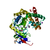
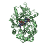
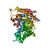
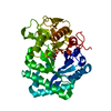
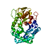
| Title | Structure of Caldicellulosiruptor danielii GH10 module of glycoside hydrolase WP_045175321 | ||||||
 Components Components | glycoside hydrolase WP_045175321 | ||||||
 Keywords Keywords | HYDROLASE / GH10 / glycoside hydrolase | ||||||
| Function / homology |  Function and homology information Function and homology informationendo-1,4-beta-xylanase activity / endo-1,4-beta-xylanase / polysaccharide catabolic process Similarity search - Function | ||||||
| Biological species |  Caldicellulosiruptor sp. Wai35.B1 (bacteria) Caldicellulosiruptor sp. Wai35.B1 (bacteria) | ||||||
| Method |  X-RAY DIFFRACTION / MOLECULAR REPLACEMENT / Resolution: 1.9 Å X-RAY DIFFRACTION / MOLECULAR REPLACEMENT / Resolution: 1.9 Å | ||||||
 Authors Authors | Alahuhta, P.M. / Lunin, V.V. | ||||||
| Funding support |  United States, 1items United States, 1items
| ||||||
 Citation Citation | Journal: AIChE J. / Year: 2019 Title: Novel multidomain, multifunctional glycoside hydrolases from highly lignocellulolytic Caldicellulosiruptor species Authors: Conway, J.M. / Crosby, J.R. / Hren, A.P. / Southerland, R.T. / Lee, L.L. / Lunin, V.V. / Alahuhta, P.M. / Himmel, M.E. / Bomble, Y.J. / Adams, M.W.W. / Kelly, R.M. | ||||||
| History |
|
- Structure visualization
| Structure viewer | Molecule: MolmilJmol/JSmol |
|---|
- Downloads & links
-Download
| PDBx/mmCIF format | 6d5c.cif.gz | 257.8 KB | Display | PDBx/mmCIF format |
|---|---|---|---|---|
| PDB format | pdb6d5c.ent.gz | 207.4 KB | Display | PDB format |
| PDBx/mmJSON format | 6d5c.json.gz | Tree view | PDBx/mmJSON format | |
| Others |  Other downloads Other downloads |
-Validation report
| Arichive directory | https://data.pdbj.org/pub/pdb/validation_reports/d5/6d5cftp://data.pdbj.org/pub/pdb/validation_reports/d5/6d5c | HTTPS FTP |
|---|
-Related structure data
| Related structure data |  6d5bC 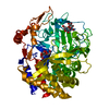 6d5dC  5ay7S S: Starting model for refinement C: citing same article ( |
|---|---|
| Similar structure data |


-Links
 PDBj
PDBj
- Assembly
| Deposited unit | 
| ||||||||
|---|---|---|---|---|---|---|---|---|---|
| 1 | 
| ||||||||
| 2 | 
| ||||||||
| 3 | 
| ||||||||
| Unit cell |
|
-Components
-Protein , 1 types, 3 molecules ABC
| #1: Protein | Mass: 40651.699 Da / Num. of mol.: 3 / Fragment: GH10 module Source method: isolated from a genetically manipulated source Source: (gene. exp.) Caldicellulosiruptor sp. Wai35.B1 (bacteria)Production host: References: UniProt: A0A4V8GZQ8*PLUS, Hydrolases; Glycosylases; Glycosidases, i.e. enzymes that hydrolyse O- and S-glycosyl compounds |
|---|
-Non-polymers , 5 types, 1313 molecules 


| #2: Chemical | ChemComp-SO4 /  Mass: 96.063 Da / Num. of mol.: 19 / Source method: obtained synthetically / Formula: SO4 Mass: 96.063 Da / Num. of mol.: 19 / Source method: obtained synthetically / Formula: SO4#3: Chemical | ChemComp-EDO /  Mass: 62.068 Da / Num. of mol.: 7 / Source method: obtained synthetically / Formula: C2H6O2 Mass: 62.068 Da / Num. of mol.: 7 / Source method: obtained synthetically / Formula: C2H6O2#4: Chemical | ChemComp-GOL /  Mass: 92.094 Da / Num. of mol.: 5 / Source method: obtained synthetically / Formula: C3H8O3 Mass: 92.094 Da / Num. of mol.: 5 / Source method: obtained synthetically / Formula: C3H8O3#5: Chemical | ChemComp-FMT / |  Mass: 46.025 Da / Num. of mol.: 1 / Source method: obtained synthetically / Formula: CH2O2 Mass: 46.025 Da / Num. of mol.: 1 / Source method: obtained synthetically / Formula: CH2O2#6: Water | ChemComp-HOH / | Mass: 18.015 Da / Num. of mol.: 1281 / Source method: isolated from a natural source / Formula: H2O |
|---|
-Experimental details
-Experiment
| Experiment | Method: X-RAY DIFFRACTION / Number of used crystals: 1 |
|---|
- Sample preparation
| Crystal | Density Matthews: 2.75 Å3/Da / Density % sol: 55.31 % |
|---|---|
| Crystal grow | Temperature: 293 K / Method: vapor diffusion, sitting drop / Details: 0.1 M Tris, pH 8-9, 1.5-2.1 M ammonium sulfate |
-Data collection
| Diffraction | Mean temperature: 100 K |
|---|---|
| Diffraction source | Source: ROTATING ANODE / Type: BRUKER AXS MICROSTAR / Wavelength: 1.5418 Å |
| Detector | Type: Bruker Platinum 135 / Detector: CCD / Date: May 31, 2016 |
| Radiation | Protocol: SINGLE WAVELENGTH / Monochromatic (M) / Laue (L): M / Scattering type: x-ray |
| Radiation wavelength | Wavelength: 1.5418 Å / Relative weight: 1 |
| Reflection | Resolution: 1.9→50 Å / Num. obs: 104001 / % possible obs: 100 % / Redundancy: 6.03 % / Rsym value: 0.068 / Net I/σ(I): 14.32 |
| Reflection shell | Resolution: 1.9→2 Å / Redundancy: 3.54 % / Mean I/σ(I) obs: 2.11 / Num. unique obs: 14807 / Rsym value: 0.4469 / % possible all: 100 |
- Processing
| Software |
| ||||||||||||||||||||||||||||||||||||||||||||||||||||||||||||||||||||||||||||||||||||||||||||||||||||||||||||||||||||||||||||||||||||||||||||||||||||||||||||||||||||||||||||||||||||||
|---|---|---|---|---|---|---|---|---|---|---|---|---|---|---|---|---|---|---|---|---|---|---|---|---|---|---|---|---|---|---|---|---|---|---|---|---|---|---|---|---|---|---|---|---|---|---|---|---|---|---|---|---|---|---|---|---|---|---|---|---|---|---|---|---|---|---|---|---|---|---|---|---|---|---|---|---|---|---|---|---|---|---|---|---|---|---|---|---|---|---|---|---|---|---|---|---|---|---|---|---|---|---|---|---|---|---|---|---|---|---|---|---|---|---|---|---|---|---|---|---|---|---|---|---|---|---|---|---|---|---|---|---|---|---|---|---|---|---|---|---|---|---|---|---|---|---|---|---|---|---|---|---|---|---|---|---|---|---|---|---|---|---|---|---|---|---|---|---|---|---|---|---|---|---|---|---|---|---|---|---|---|---|---|
| Refinement | Method to determine structure: MOLECULAR REPLACEMENT Starting model: PDB entry 5AY7 Resolution: 1.9→86.48 Å / Cor.coef. Fo:Fc: 0.966 / Cor.coef. Fo:Fc free: 0.938 / SU B: 4.029 / SU ML: 0.113 / Cross valid method: THROUGHOUT / ESU R: 0.144 / ESU R Free: 0.144 / Details: HYDROGENS HAVE BEEN ADDED IN THE RIDING POSITIONS
| ||||||||||||||||||||||||||||||||||||||||||||||||||||||||||||||||||||||||||||||||||||||||||||||||||||||||||||||||||||||||||||||||||||||||||||||||||||||||||||||||||||||||||||||||||||||
| Solvent computation | Ion probe radii: 0.8 Å / Shrinkage radii: 0.8 Å / VDW probe radii: 1.2 Å | ||||||||||||||||||||||||||||||||||||||||||||||||||||||||||||||||||||||||||||||||||||||||||||||||||||||||||||||||||||||||||||||||||||||||||||||||||||||||||||||||||||||||||||||||||||||
| Displacement parameters | Biso mean: 27.186 Å2
| ||||||||||||||||||||||||||||||||||||||||||||||||||||||||||||||||||||||||||||||||||||||||||||||||||||||||||||||||||||||||||||||||||||||||||||||||||||||||||||||||||||||||||||||||||||||
| Refinement step | Cycle: 1 / Resolution: 1.9→86.48 Å
| ||||||||||||||||||||||||||||||||||||||||||||||||||||||||||||||||||||||||||||||||||||||||||||||||||||||||||||||||||||||||||||||||||||||||||||||||||||||||||||||||||||||||||||||||||||||
| Refine LS restraints |
|