 Movie
Movie Controller


+ Open data

- Basic information
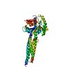
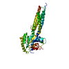
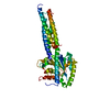
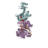
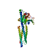
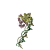
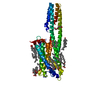
| Entry | Database: PDB / ID: 6cc7 | |||||||||
|---|---|---|---|---|---|---|---|---|---|---|
| Title | Structure of Fungal GH62 from Thielavia terretris | |||||||||
 Components Components | Glycoside hydrolase family 62 protein | |||||||||
 Keywords Keywords |  HYDROLASE / GH62 / glycoside hydrolase / domain swapping / Thielavia terrestris HYDROLASE / GH62 / glycoside hydrolase / domain swapping / Thielavia terrestris | |||||||||
| Function / homology | Glycoside hydrolase, family 62, arabinosidase / Glycosyl hydrolase family 62 / L-arabinose metabolic process / non-reducing end alpha-L-arabinofuranosidase / alpha-L-arabinofuranosidase activity / Glycosyl hydrolase, five-bladed beta-propellor domain superfamily / xylan catabolic process / extracellular region / Non-reducing end alpha-L-arabinofuranosidase Function and homology information Function and homology information | |||||||||
| Biological species |  Thielavia terrestris (fungus) Thielavia terrestris (fungus) | |||||||||
| Method | X-RAY DIFFRACTION / SYNCHROTRON / MOLECULAR REPLACEMENT / Resolution: 1.87 Å | |||||||||
 Authors Authors | Camargo, S. / Mulinari, E.J. / Almeida, L.R. / Muniz, J.R.C. | |||||||||
| Funding support |  Brazil, 2items Brazil, 2items
| |||||||||
 Citation Citation | Journal: To Be Published Title: Structure of Fungal GH62 from Thielavia terretris Authors: Camargo, S. / Mulinari, E.J. / Almeida, L.R. / Muniz, J.R.C. | |||||||||
| History |
|
- Structure visualization
| Structure viewer | Molecule: MolmilJmol/JSmol |
|---|
- Downloads & links
-Download
| PDBx/mmCIF format | 6cc7.cif.gz | 133.6 KB | Display | PDBx/mmCIF format |
|---|---|---|---|---|
| PDB format | pdb6cc7.ent.gz | 102.7 KB | Display | PDB format |
| PDBx/mmJSON format | 6cc7.json.gz | Tree view | PDBx/mmJSON format | |
| Others |  Other downloads Other downloads |
-Validation report
| Arichive directory | https://data.pdbj.org/pub/pdb/validation_reports/cc/6cc7ftp://data.pdbj.org/pub/pdb/validation_reports/cc/6cc7 | HTTPS FTP |
|---|
-Related structure data
| Related structure data |  4o8nS S: Starting model for refinement |
|---|---|
| Similar structure data |
-Links
 PDBj
PDBj- Assembly
| Deposited unit | 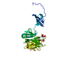
| |||||||||||||||
|---|---|---|---|---|---|---|---|---|---|---|---|---|---|---|---|---|
| 1 |
| |||||||||||||||
| Unit cell |
| |||||||||||||||
| Components on special symmetry positions |
|
-Components
| #1: Protein | Mass: 34801.578 Da / Num. of mol.: 1 Source method: isolated from a genetically manipulated source Source: (gene. exp.) Thielavia terrestris (fungus) / Strain: ATCC 38088 / NRRL 8126 / Gene: THITE_2110954 / Production host:  Escherichia coli (E. coli) / References: UniProt: G2QVH2 Escherichia coli (E. coli) / References: UniProt: G2QVH2 | ||||
|---|---|---|---|---|---|
| #2: Chemical | ChemComp-CA /   Mass: 40.078 Da / Num. of mol.: 1 / Source method: obtained synthetically / Formula: Ca Mass: 40.078 Da / Num. of mol.: 1 / Source method: obtained synthetically / Formula: Ca | ||||
| #3: Chemical | Sulfate  Mass: 96.063 Da / Num. of mol.: 2 / Source method: obtained synthetically / Formula: SO4 Mass: 96.063 Da / Num. of mol.: 2 / Source method: obtained synthetically / Formula: SO4#4: Chemical | ChemComp-EDO / | Ethylene glycol  Mass: 62.068 Da / Num. of mol.: 1 / Source method: obtained synthetically / Formula: C2H6O2 Mass: 62.068 Da / Num. of mol.: 1 / Source method: obtained synthetically / Formula: C2H6O2#5: Water | ChemComp-HOH / | Water Mass: 18.015 Da / Num. of mol.: 298 / Source method: isolated from a natural source / Formula: H2O Mass: 18.015 Da / Num. of mol.: 298 / Source method: isolated from a natural source / Formula: H2O |
-Experimental details
-Experiment
| Experiment | Method: X-RAY DIFFRACTION / Number of used crystals: 1 |
|---|
- Sample preparation
| Crystal | Density Matthews: 1.89 Å3/Da / Density % sol: 31.2 % |
|---|---|
| Crystal grow | Temperature: 291 K / Method: vapor diffusion, sitting drop / pH: 4.6 / Details: 0.1 M sodium acetate, 2 M ammonium sulfate |
-Data collection
| Diffraction | Mean temperature: 100 K |
|---|---|
| Diffraction source | Source: SYNCHROTRON / Site: LNLS / Beamline: W01B-MX2 / Wavelength: 1.45863 Å |
| Detector | Type: DECTRIS PILATUS 2M / Detector: PIXEL / Date: Oct 3, 2016 |
| Radiation | Protocol: SINGLE WAVELENGTH / Monochromatic (M) / Laue (L): M / Scattering type: x-ray |
| Radiation wavelength | Wavelength: 1.45863 Å / Relative weight: 1 |
| Reflection | Resolution: 1.87→57.48 Å / Num. obs: 20946 / % possible obs: 99.8 % / Observed criterion σ(I): 2 / Redundancy: 5.5 % / CC1/2: 0.998 / Rmerge(I) obs: 0.071 / Rpim(I) all: 0.045 / Rrim(I) all: 0.086 / Net I/σ(I): 11.9 |
| Reflection shell | Resolution: 1.87→1.91 Å / Redundancy: 2.8 % / Rmerge(I) obs: 0.386 / Mean I/σ(I) obs: 2.2 / Num. unique obs: 1318 / CC1/2: 0.838 / Rpim(I) all: 0.349 / Rrim(I) all: 0.522 / % possible all: 97.4 |
- Processing
| Software |
| ||||||||||||||||||||||||||||||||||||||||||||||||||||||||||||||||||||||||||||||||||||||||||||||||||||||||||||||||||||||||||||||||||||||||||||||||||||||||||||||||||||||||||||||||||||||
|---|---|---|---|---|---|---|---|---|---|---|---|---|---|---|---|---|---|---|---|---|---|---|---|---|---|---|---|---|---|---|---|---|---|---|---|---|---|---|---|---|---|---|---|---|---|---|---|---|---|---|---|---|---|---|---|---|---|---|---|---|---|---|---|---|---|---|---|---|---|---|---|---|---|---|---|---|---|---|---|---|---|---|---|---|---|---|---|---|---|---|---|---|---|---|---|---|---|---|---|---|---|---|---|---|---|---|---|---|---|---|---|---|---|---|---|---|---|---|---|---|---|---|---|---|---|---|---|---|---|---|---|---|---|---|---|---|---|---|---|---|---|---|---|---|---|---|---|---|---|---|---|---|---|---|---|---|---|---|---|---|---|---|---|---|---|---|---|---|---|---|---|---|---|---|---|---|---|---|---|---|---|---|---|
| Refinement | Method to determine structure: MOLECULAR REPLACEMENT Starting model: 4o8n Resolution: 1.87→57.48 Å / Cor.coef. Fo:Fc: 0.974 / Cor.coef. Fo:Fc free: 0.952 / SU B: 6.634 / SU ML: 0.101 / Cross valid method: THROUGHOUT / ESU R: 0.151 / ESU R Free: 0.136 / Stereochemistry target values: MAXIMUM LIKELIHOOD / Details: HYDROGENS HAVE BEEN ADDED IN THE RIDING POSITIONS
| ||||||||||||||||||||||||||||||||||||||||||||||||||||||||||||||||||||||||||||||||||||||||||||||||||||||||||||||||||||||||||||||||||||||||||||||||||||||||||||||||||||||||||||||||||||||
| Solvent computation | Ion probe radii: 0.8 Å / Shrinkage radii: 0.8 Å / VDW probe radii: 1.2 Å / Solvent model: MASK | ||||||||||||||||||||||||||||||||||||||||||||||||||||||||||||||||||||||||||||||||||||||||||||||||||||||||||||||||||||||||||||||||||||||||||||||||||||||||||||||||||||||||||||||||||||||
| Displacement parameters | Biso mean: 23.69 Å2
| ||||||||||||||||||||||||||||||||||||||||||||||||||||||||||||||||||||||||||||||||||||||||||||||||||||||||||||||||||||||||||||||||||||||||||||||||||||||||||||||||||||||||||||||||||||||
| Refinement step | Cycle: 1 / Resolution: 1.87→57.48 Å
| ||||||||||||||||||||||||||||||||||||||||||||||||||||||||||||||||||||||||||||||||||||||||||||||||||||||||||||||||||||||||||||||||||||||||||||||||||||||||||||||||||||||||||||||||||||||
| Refine LS restraints |
|