DRM complex / Myb complex / TFAP2A acts as a transcriptional repressor during retinoic acid induced cell differentiation / Transcription of E2F targets under negative control by p107 (RBL1) and p130 (RBL2) in complex with HDAC1 / Polo-like kinase mediated events / Transcription of E2F targets under negative control by DREAM complex / DNA biosynthetic process / G1/S-Specific Transcription / G0 and Early G1 / mitotic spindle assembly ...DRM complex / Myb complex / TFAP2A acts as a transcriptional repressor during retinoic acid induced cell differentiation / Transcription of E2F targets under negative control by p107 (RBL1) and p130 (RBL2) in complex with HDAC1 / Polo-like kinase mediated events / Transcription of E2F targets under negative control by DREAM complex / DNA biosynthetic process / G1/S-Specific Transcription / G0 and Early G1 / mitotic spindle assembly / Cyclin E associated events during G1/S transition / Cyclin A:Cdk2-associated events at S phase entry / transcription repressor complex / cellular response to leukemia inhibitory factor / sequence-specific double-stranded DNA binding / mitotic cell cycle / positive regulation of neuron apoptotic process / DNA-binding transcription activator activity, RNA polymerase II-specific / DNA-binding transcription factor activity, RNA polymerase II-specific / regulation of cell cycle / RNA polymerase II cis-regulatory region sequence-specific DNA binding / regulation of transcription by RNA polymerase II / regulation of DNA-templated transcription / DNA-templated transcription / positive regulation of transcription by RNA polymerase II / DNA binding / nucleoplasm / nucleus / cytosol Similarity search - Function
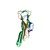
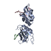
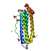
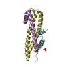
A: Protein lin-9 homolog D: Protein lin-9 homolog F: Myb-related protein B C: Myb-related protein B B: Protein lin-52 homolog E: Protein lin-52 homolog hetero molecules
Proteinlin-9homolog / hLin-9 / Beta subunit-associated regulator of apoptosis / TUDOR gene similar protein / Type I ...hLin-9 / Beta subunit-associated regulator of apoptosis / TUDOR gene similar protein / Type I interferon receptor beta chain-associated protein / pRB-associated protein
Mass: 13999.179 Da / Num. of mol.: 2 Source method: isolated from a genetically manipulated source Source: (gene. exp.) Homo sapiens (human) / Gene: LIN9, BARA, TGS / Production host: Escherichia coli (E. coli) / References: UniProt: Q5TKA1
#2: Protein/peptide
Myb-relatedproteinB / B-Myb / Myb-like protein 2
Mass: 3649.959 Da / Num. of mol.: 2 Source method: isolated from a genetically manipulated source Source: (gene. exp.) Homo sapiens (human) / Gene: MYBL2, BMYB / Production host: Escherichia coli (E. coli) / References: UniProt: P10244
#3: Protein
Proteinlin-52homolog
Mass: 7823.031 Da / Num. of mol.: 2 Source method: isolated from a genetically manipulated source Source: (gene. exp.) Homo sapiens (human) / Gene: LIN52, C14orf46 / Production host: Escherichia coli (E. coli) / References: UniProt: Q52LA3
In the structure databanks used in Yorodumi, some data are registered as the other names, "COVID-19 virus" and "2019-nCoV". Here are the details of the virus and the list of structure data.
Jan 31, 2019. EMDB accession codes are about to change! (news from PDBe EMDB page)
EMDB accession codes are about to change! (news from PDBe EMDB page)
The allocation of 4 digits for EMDB accession codes will soon come to an end. Whilst these codes will remain in use, new EMDB accession codes will include an additional digit and will expand incrementally as the available range of codes is exhausted. The current 4-digit format prefixed with “EMD-” (i.e. EMD-XXXX) will advance to a 5-digit format (i.e. EMD-XXXXX), and so on. It is currently estimated that the 4-digit codes will be depleted around Spring 2019, at which point the 5-digit format will come into force.
The EM Navigator/Yorodumi systems omit the EMD- prefix.
Related info.:Q: What is EMD? / ID/Accession-code notation in Yorodumi/EM Navigator
Yorodumi is a browser for structure data from EMDB, PDB, SASBDB, etc.
This page is also the successor to EM Navigator detail page, and also detail information page/front-end page for Omokage search.
The word "yorodu" (or yorozu) is an old Japanese word meaning "ten thousand". "mi" (miru) is to see.
Related info.:EMDB / PDB / SASBDB / Comparison of 3 databanks / Yorodumi Search / Aug 31, 2016. New EM Navigator & Yorodumi / Yorodumi Papers / Jmol/JSmol / Function and homology information / Changes in new EM Navigator and Yorodumi
 Movie
Movie Controller


 Open data
 Basic information
 Components
 Keywords
 Function and homology information
 Homo sapiens (human)
 X-RAY DIFFRACTION /
  Authors
 United States, 2items
  Citation
 Structure visualization
 Downloads & links
 Other downloads
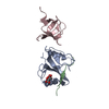


 PDBj
PDBj


 Assembly


 Mass: 96.063 Da / Num. of mol.: 2 / Source method: obtained synthetically / Formula: SO4
 Mass: 18.015 Da / Num. of mol.: 83 / Source method: isolated from a natural source / Formula: H2O
 Sample preparation
 Processing