 Movie
Movie Controller


+ Open data

- Basic information
| Entry | Database: PDB / ID: 4hn7 | ||||||
|---|---|---|---|---|---|---|---|
| Title | Crystal structure of E. coli PmrD | ||||||
 Components Components | Signal transduction protein pmrD | ||||||
 Keywords Keywords | SIGNALING PROTEIN | ||||||
| Function / homology | Signal transduction protein PmrD / IraM/PmrD / OB fold (Dihydrolipoamide Acetyltransferase, E2P) - #650 / Signal transduction protein PmrD superfamily / OB fold (Dihydrolipoamide Acetyltransferase, E2P) / response to antibiotic / Beta Barrel / Mainly Beta / Signal transduction protein PmrD Function and homology information Function and homology information | ||||||
| Biological species |  | ||||||
| Method |  X-RAY DIFFRACTION / SYNCHROTRON / MOLECULAR REPLACEMENT / Resolution: 2.352 Å X-RAY DIFFRACTION / SYNCHROTRON / MOLECULAR REPLACEMENT / Resolution: 2.352 Å | ||||||
 Authors Authors | Jeong, E. / Jung, H. / Ban, C. | ||||||
 Citation Citation | Journal: To be Published Title: Crystal structure of E. coli PmrD Authors: Jeong, E. / Jung, H. / Ban, C. | ||||||
| History |
|
- Structure visualization
| Structure viewer | Molecule: MolmilJmol/JSmol |
|---|
- Downloads & links
-Download
| PDBx/mmCIF format | 4hn7.cif.gz | 29 KB | Display | PDBx/mmCIF format |
|---|---|---|---|---|
| PDB format | pdb4hn7.ent.gz | 18.4 KB | Display | PDB format |
| PDBx/mmJSON format | 4hn7.json.gz | Tree view | PDBx/mmJSON format | |
| Others |  Other downloads Other downloads |
-Validation report
| Arichive directory | https://data.pdbj.org/pub/pdb/validation_reports/hn/4hn7ftp://data.pdbj.org/pub/pdb/validation_reports/hn/4hn7 | HTTPS FTP |
|---|
-Related structure data
| Related structure data |  2jsoS S: Starting model for refinement |
|---|---|
| Similar structure data |


-Links
 PDBj
PDBj- Assembly
| Deposited unit | 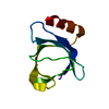
| ||||||||
|---|---|---|---|---|---|---|---|---|---|
| 1 | 
| ||||||||
| Unit cell |
|
-Components
| #1: Protein | Mass: 9885.536 Da / Num. of mol.: 1 Source method: isolated from a genetically manipulated source Source: (gene. exp.) |
|---|---|
| #2: Water | ChemComp-HOH /  Mass: 18.015 Da / Num. of mol.: 7 / Source method: isolated from a natural source / Formula: H2O Mass: 18.015 Da / Num. of mol.: 7 / Source method: isolated from a natural source / Formula: H2O |
| Has protein modification | Y |
-Experimental details
-Experiment
| Experiment | Method: X-RAY DIFFRACTION / Number of used crystals: 1 |
|---|
- Sample preparation
| Crystal | Density Matthews: 2.58 Å3/Da / Density % sol: 52.37 % |
|---|---|
| Crystal grow | Temperature: 293 K / Method: vapor diffusion, hanging drop / pH: 4.6 Details: 50 mM sodium acetate, 2.0 M ammonium sulfate, pH 4.6, VAPOR DIFFUSION, HANGING DROP, temperature 293.0K |
-Data collection
| Diffraction | Mean temperature: 100 K | ||||||||||||
|---|---|---|---|---|---|---|---|---|---|---|---|---|---|
| Diffraction source | Source: SYNCHROTRON / Site: PAL/PLS  / Beamline: 4A / Wavelength: 0.97959 Å / Beamline: 4A / Wavelength: 0.97959 Å | ||||||||||||
| Detector | Type: ADSC QUANTUM 315 / Detector: CCD / Date: Jul 22, 2005 / Details: mirrors | ||||||||||||
| Radiation | Monochromator: Double crystal monochromator / Protocol: MAD / Monochromatic (M) / Laue (L): M / Scattering type: x-ray | ||||||||||||
| Radiation wavelength | Wavelength: 0.97959 Å / Relative weight: 1 | ||||||||||||
| Reflection | Resolution: 2→50 Å / Num. obs: 5617 / % possible obs: 79.6 % / Observed criterion σ(F): 0 / Observed criterion σ(I): -3 | ||||||||||||
| Reflection shell |
|
- Processing
| Software |
| ||||||||||||||||||||||||||||||||||||||||||||||||||||||||||||||||||||||||||||||||||||||||||||||||||||||||||||||||||||||||||||||||||||||||||||||||||||||||||||||||||||||||||
|---|---|---|---|---|---|---|---|---|---|---|---|---|---|---|---|---|---|---|---|---|---|---|---|---|---|---|---|---|---|---|---|---|---|---|---|---|---|---|---|---|---|---|---|---|---|---|---|---|---|---|---|---|---|---|---|---|---|---|---|---|---|---|---|---|---|---|---|---|---|---|---|---|---|---|---|---|---|---|---|---|---|---|---|---|---|---|---|---|---|---|---|---|---|---|---|---|---|---|---|---|---|---|---|---|---|---|---|---|---|---|---|---|---|---|---|---|---|---|---|---|---|---|---|---|---|---|---|---|---|---|---|---|---|---|---|---|---|---|---|---|---|---|---|---|---|---|---|---|---|---|---|---|---|---|---|---|---|---|---|---|---|---|---|---|---|---|---|---|---|---|---|
| Refinement | Method to determine structure: MOLECULAR REPLACEMENT Starting model: PDB ENTRY 2JSO Resolution: 2.352→16.81 Å / Cor.coef. Fo:Fc: 0.958 / Cor.coef. Fo:Fc free: 0.93 / SU B: 10.697 / SU ML: 0.227 / Cross valid method: THROUGHOUT / σ(F): 0 / ESU R: 0.346 / ESU R Free: 0.273 / Stereochemistry target values: MAXIMUM LIKELIHOOD / Details: HYDROGENS HAVE BEEN ADDED IN THE RIDING POSITIONS
| ||||||||||||||||||||||||||||||||||||||||||||||||||||||||||||||||||||||||||||||||||||||||||||||||||||||||||||||||||||||||||||||||||||||||||||||||||||||||||||||||||||||||||
| Solvent computation | Ion probe radii: 0.8 Å / Shrinkage radii: 0.8 Å / VDW probe radii: 1.2 Å / Solvent model: MASK | ||||||||||||||||||||||||||||||||||||||||||||||||||||||||||||||||||||||||||||||||||||||||||||||||||||||||||||||||||||||||||||||||||||||||||||||||||||||||||||||||||||||||||
| Displacement parameters | Biso mean: 74.083 Å2
| ||||||||||||||||||||||||||||||||||||||||||||||||||||||||||||||||||||||||||||||||||||||||||||||||||||||||||||||||||||||||||||||||||||||||||||||||||||||||||||||||||||||||||
| Refinement step | Cycle: LAST / Resolution: 2.352→16.81 Å
| ||||||||||||||||||||||||||||||||||||||||||||||||||||||||||||||||||||||||||||||||||||||||||||||||||||||||||||||||||||||||||||||||||||||||||||||||||||||||||||||||||||||||||
| Refine LS restraints |
| ||||||||||||||||||||||||||||||||||||||||||||||||||||||||||||||||||||||||||||||||||||||||||||||||||||||||||||||||||||||||||||||||||||||||||||||||||||||||||||||||||||||||||
| LS refinement shell | Resolution: 2.352→2.412 Å / Total num. of bins used: 20
|
