 Movie
Movie Controller

[English] 日本語
 Yorodumi
- PDB-6a68: the crystal structure of rat calcium-dependent activator protein ... -
+ Open data

- Basic information
| Entry | Database: PDB / ID: 6a68 | ||||||
|---|---|---|---|---|---|---|---|
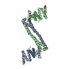
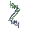
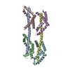
| Title | the crystal structure of rat calcium-dependent activator protein for secretion (CAPS) DAMH domain | ||||||
 Components Components | Calcium-dependent secretion activator 1 | ||||||
 Keywords Keywords |  EXOCYTOSIS / exocytosis DCV transition EXOCYTOSIS / exocytosis DCV transition | ||||||
| Function / homology |  Function and homology informationextrinsic component of neuronal dense core vesicle membrane / catecholamine secretion / dense core granule / regulated exocytosis / presynaptic dense core vesicle exocytosis / vesicle organization / positive regulation of calcium ion-dependent exocytosis / synaptic vesicle priming / exocytosis / positive regulation of exocytosis ...extrinsic component of neuronal dense core vesicle membrane / catecholamine secretion / dense core granule / regulated exocytosis / presynaptic dense core vesicle exocytosis / vesicle organization / positive regulation of calcium ion-dependent exocytosis / synaptic vesicle priming / exocytosis / positive regulation of exocytosis / phosphatidylinositol-4,5-bisphosphate binding / establishment of localization in cell / protein transport / presynapse / cytoplasmic vesicle / glutamatergic synapse / calcium ion binding / protein kinase binding Function and homology informationextrinsic component of neuronal dense core vesicle membrane / catecholamine secretion / dense core granule / regulated exocytosis / presynaptic dense core vesicle exocytosis / vesicle organization / positive regulation of calcium ion-dependent exocytosis / synaptic vesicle priming / exocytosis / positive regulation of exocytosis ...extrinsic component of neuronal dense core vesicle membrane / catecholamine secretion / dense core granule / regulated exocytosis / presynaptic dense core vesicle exocytosis / vesicle organization / positive regulation of calcium ion-dependent exocytosis / synaptic vesicle priming / exocytosis / positive regulation of exocytosis / phosphatidylinositol-4,5-bisphosphate binding / establishment of localization in cell / protein transport / presynapse / cytoplasmic vesicle / glutamatergic synapse / calcium ion binding / protein kinase bindingSimilarity search - Function | ||||||
| Biological species |  Rattus norvegicus (Norway rat) Rattus norvegicus (Norway rat) | ||||||
| Method | X-RAY DIFFRACTION / SYNCHROTRON / SAD / Resolution: 2.901 Å | ||||||
 Authors Authors | Zhou, H. / Wei, Z.Q. / Yao, D.Q. / Zhang, R.G. / Ma, C. | ||||||
 Citation Citation | Journal: Cell Rep / Year: 2019 Title: Structural and Functional Analysis of the CAPS SNARE-Binding Domain Required for SNARE Complex Formation and Exocytosis. Authors: Zhou, H. / Wei, Z. / Wang, S. / Yao, D. / Zhang, R. / Ma, C. | ||||||
| History |
|
- Structure visualization
| Structure viewer | Molecule: MolmilJmol/JSmol |
|---|
- Downloads & links
-Download
| PDBx/mmCIF format | 6a68.cif.gz | 43.9 KB | Display | PDBx/mmCIF format |
|---|---|---|---|---|
| PDB format | pdb6a68.ent.gz | 32.1 KB | Display | PDB format |
| PDBx/mmJSON format | 6a68.json.gz | Tree view | PDBx/mmJSON format | |
| Others |  Other downloads Other downloads |
-Validation report
| Arichive directory | https://data.pdbj.org/pub/pdb/validation_reports/a6/6a68ftp://data.pdbj.org/pub/pdb/validation_reports/a6/6a68 | HTTPS FTP |
|---|
-Related structure data
| Similar structure data |
|---|


-Links
 PDBj
PDBj


- Assembly
| Deposited unit | 
| ||||||||
|---|---|---|---|---|---|---|---|---|---|
| 1 | 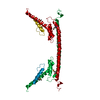
| ||||||||
| Unit cell |
| ||||||||
| Components on special symmetry positions |
|
-Components
| #1: Protein | Mass: 21562.670 Da / Num. of mol.: 1 Source method: isolated from a genetically manipulated source Source: (gene. exp.) Rattus norvegicus (Norway rat) / Gene: Cadps, Caps, Caps1 / Production host:  Escherichia coli K-12 (bacteria) / References: UniProt: Q62717 Escherichia coli K-12 (bacteria) / References: UniProt: Q62717 |
|---|---|
| #2: Chemical | ChemComp-K /   Mass: 39.098 Da / Num. of mol.: 1 / Source method: obtained synthetically / Formula: K Mass: 39.098 Da / Num. of mol.: 1 / Source method: obtained synthetically / Formula: K |
| #3: Water | ChemComp-HOH / Water Mass: 18.015 Da / Num. of mol.: 11 / Source method: isolated from a natural source / Formula: H2O Mass: 18.015 Da / Num. of mol.: 11 / Source method: isolated from a natural source / Formula: H2O |
-Experimental details
-Experiment
| Experiment | Method: X-RAY DIFFRACTION / Number of used crystals: 1 |
|---|
- Sample preparation
| Crystal | Density Matthews: 6.13 Å3/Da / Density % sol: 79.93 % |
|---|---|
| Crystal grow | Temperature: 293 K / Method: vapor diffusion, sitting drop Details: 18-25% (v/v) PEG 3350, 0.1M Succinic acid (pH 7.0-7.5) |
-Data collection
| Diffraction | Mean temperature: 100 K |
|---|---|
| Diffraction source | Source: SYNCHROTRON / Site: SSRF  / Beamline: BL18U1 / Wavelength: 0.9793 Å / Beamline: BL18U1 / Wavelength: 0.9793 Å |
| Detector | Type: ADSC QUANTUM 315r / Detector: CCD / Date: Mar 12, 2017 |
| Radiation | Protocol: SINGLE WAVELENGTH / Monochromatic (M) / Laue (L): M / Scattering type: x-ray |
| Radiation wavelength | Wavelength: 0.9793 Å / Relative weight: 1 |
| Reflection | Resolution: 2.9→25 Å / Num. obs: 12274 / % possible obs: 99.7 % / Redundancy: 9.6 % / Rmerge(I) obs: 0.127 / Rpim(I) all: 0.041 / Net I/σ(I): 16.8 |
| Reflection shell | Resolution: 2.9→2.95 Å / Redundancy: 9.2 % / Rmerge(I) obs: 0.828 / Mean I/σ(I) obs: 2.4 / Rpim(I) all: 0.276 / % possible all: 99.8 |
-Phasing
| Phasing | Method: SAD |
|---|
- Processing
| Software |
| |||||||||||||||||||||||||||||||||||
|---|---|---|---|---|---|---|---|---|---|---|---|---|---|---|---|---|---|---|---|---|---|---|---|---|---|---|---|---|---|---|---|---|---|---|---|---|
| Refinement | Method to determine structure: SAD / Resolution: 2.901→24.884 Å / SU ML: 0.46 / Cross valid method: THROUGHOUT / σ(F): 1.36 / Phase error: 29.19 / Stereochemistry target values: ML Details: Anomalous signal for residue 0 MET was very weak, and could not be identified as MSE.
| |||||||||||||||||||||||||||||||||||
| Solvent computation | Shrinkage radii: 0.9 Å / VDW probe radii: 1.11 Å / Solvent model: FLAT BULK SOLVENT MODEL | |||||||||||||||||||||||||||||||||||
| Displacement parameters | Biso max: 153.88 Å2 / Biso mean: 49.1338 Å2 / Biso min: 6.4 Å2 | |||||||||||||||||||||||||||||||||||
| Refinement step | Cycle: final / Resolution: 2.901→24.884 Å
| |||||||||||||||||||||||||||||||||||
| Refine LS restraints |
| |||||||||||||||||||||||||||||||||||
| LS refinement shell | Refine-ID: X-RAY DIFFRACTION / Rfactor Rfree error: 0 / Total num. of bins used: 4
|
