 Movie
Movie Controller


+ Open data

- Basic information
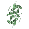
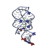
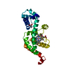
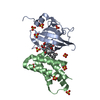
| Entry | Database: PDB / ID: 5t1i | ||||||
|---|---|---|---|---|---|---|---|
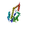
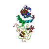
| Title | CBX3 chromo shadow domain in complex with histone H3 peptide | ||||||
 Components Components |
| ||||||
 Keywords Keywords |  TRANSCRIPTION / Structural Genomics / Structural Genomics Consortium / SGC TRANSCRIPTION / Structural Genomics / Structural Genomics Consortium / SGC | ||||||
| Function / homology |  Function and homology information Function and homology informationchromatin lock complex / histone methyltransferase binding / condensed chromosome, centromeric region / nuclear inner membrane / Transcriptional Regulation by E2F6 / site of DNA damage / chromosome, centromeric region / heterochromatin / Chromatin modifying enzymes / pericentric heterochromatin ...chromatin lock complex / histone methyltransferase binding / condensed chromosome, centromeric region / nuclear inner membrane / Transcriptional Regulation by E2F6 / site of DNA damage / chromosome, centromeric region / heterochromatin / Chromatin modifying enzymes / pericentric heterochromatin / heterochromatin formation / epigenetic regulation of gene expression / methylated histone binding / telomere organization / RNA Polymerase I Promoter Opening / Interleukin-7 signaling / Assembly of the ORC complex at the origin of replication / DNA methylation / Condensation of Prophase Chromosomes / HCMV Late Events / Chromatin modifications during the maternal to zygotic transition (MZT) / ERCC6 (CSB) and EHMT2 (G9a) positively regulate rRNA expression / SIRT1 negatively regulates rRNA expression / PRC2 methylates histones and DNA / Defective pyroptosis / HDACs deacetylate histones / transcription coregulator binding / RNA Polymerase I Promoter Escape / Transcriptional regulation by small RNAs / Formation of the beta-catenin:TCF transactivating complex / euchromatin / RUNX1 regulates genes involved in megakaryocyte differentiation and platelet function / Activated PKN1 stimulates transcription of AR (androgen receptor) regulated genes KLK2 and KLK3 / NoRC negatively regulates rRNA expression / B-WICH complex positively regulates rRNA expression / HDMs demethylate histones / spindle / PKMTs methylate histone lysines / RMTs methylate histone arginines / Meiotic recombination / Pre-NOTCH Transcription and Translation / nucleosome assembly / Activation of anterior HOX genes in hindbrain development during early embryogenesis / HCMV Early Events / Transcriptional regulation of granulopoiesis / structural constituent of chromatin / RNA polymerase II transcription regulator complex / rhythmic process / nucleosome / nuclear envelope / gene expression / RUNX1 regulates transcription of genes involved in differentiation of HSCs / chromatin organization / Factors involved in megakaryocyte development and platelet production / HATs acetylate histones / Senescence-Associated Secretory Phenotype (SASP) / Oxidative Stress Induced Senescence / Estrogen-dependent gene expression / chromosome, telomeric region / chromatin remodeling / cadherin binding / Amyloid fiber formation / protein heterodimerization activity / protein domain specific binding / negative regulation of DNA-templated transcription / DNA damage response / chromatin binding / chromatin / negative regulation of transcription by RNA polymerase II / enzyme binding / protein-containing complex / DNA binding / extracellular exosome / extracellular region / nucleoplasm / membrane / identical protein binding / nucleusSimilarity search - Function | ||||||
| Biological species |  Homo sapiens (human) Homo sapiens (human) | ||||||
| Method | X-RAY DIFFRACTION / SYNCHROTRON / MOLECULAR REPLACEMENT / Resolution: 1.6 Å | ||||||
 Authors Authors | Liu, Y. / Tempel, W. / Bountra, C. / Arrowsmith, C.H. / Edwards, A.M. / Min, J. / Structural Genomics Consortium (SGC) | ||||||
 Citation Citation | Journal: J. Biol. Chem. / Year: 2017 Title: Peptide recognition by heterochromatin protein 1 (HP1) chromoshadow domains revisited: Plasticity in the pseudosymmetric histone binding site of human HP1. Authors: Liu, Y. / Qin, S. / Lei, M. / Tempel, W. / Zhang, Y. / Loppnau, P. / Li, Y. / Min, J. | ||||||
| History |
|
- Structure visualization
| Structure viewer | Molecule: MolmilJmol/JSmol |
|---|
- Downloads & links
-Download
| PDBx/mmCIF format | 5t1i.cif.gz | 76.5 KB | Display | PDBx/mmCIF format |
|---|---|---|---|---|
| PDB format | pdb5t1i.ent.gz | 55.8 KB | Display | PDB format |
| PDBx/mmJSON format | 5t1i.json.gz | Tree view | PDBx/mmJSON format | |
| Others |  Other downloads Other downloads |
-Validation report
| Arichive directory | https://data.pdbj.org/pub/pdb/validation_reports/t1/5t1iftp://data.pdbj.org/pub/pdb/validation_reports/t1/5t1i | HTTPS FTP |
|---|
-Related structure data
| Related structure data |  3kupS S: Starting model for refinement |
|---|---|
| Similar structure data |


-Links
 PDBj
PDBj


- Assembly
| Deposited unit | 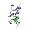
| ||||||||
|---|---|---|---|---|---|---|---|---|---|
| 1 |
| ||||||||
| Unit cell |
| ||||||||
| Details | Authors did not experimentally determine the biological unit, but assigned it homologously to PDB entry 1S4Z. |
-Components
| #1: Protein | Mass: 7637.704 Da / Num. of mol.: 2 / Fragment: unp residues 110-176 Source method: isolated from a genetically manipulated source Source: (gene. exp.) Homo sapiens (human) / Gene: CBX3 / Plasmid: pET28-MHL / Production host:  Escherichia coli (E. coli) / Strain (production host): BL21(DE3)-V2R-pRARE2 / References: UniProt: Q13185 Escherichia coli (E. coli) / Strain (production host): BL21(DE3)-V2R-pRARE2 / References: UniProt: Q13185#2: Protein/peptide | | Histone H3 / Histone H3/a / Histone H3/b / Histone H3/c / Histone H3/d / Histone H3/f / Histone H3/h / Histone ...Histone H3/a / Histone H3/b / Histone H3/c / Histone H3/d / Histone H3/f / Histone H3/h / Histone H3/i / Histone H3/j / Histone H3/k / Histone H3/lMass: 1867.186 Da / Num. of mol.: 1 / Fragment: unp residues 39-53 / Source method: obtained synthetically / Source: (synth.) Homo sapiens (human) / References: UniProt: P68431#3: Chemical | ChemComp-UNX /   Num. of mol.: 12 / Source method: obtained synthetically Num. of mol.: 12 / Source method: obtained synthetically#4: Water | ChemComp-HOH / | Water Mass: 18.015 Da / Num. of mol.: 44 / Source method: isolated from a natural source / Formula: H2O Mass: 18.015 Da / Num. of mol.: 44 / Source method: isolated from a natural source / Formula: H2O |
|---|
-Experimental details
-Experiment
| Experiment | Method: X-RAY DIFFRACTION / Number of used crystals: 1 |
|---|
- Sample preparation
| Crystal | Density Matthews: 1.9 Å3/Da / Density % sol: 35.31 % |
|---|---|
| Crystal grow | Temperature: 293 K / Method: vapor diffusion, sitting drop / pH: 7.5 Details: 25% PEG 3350, 0.2 M sodium chloride, 0.1 M Hepes, 5% Ethylene Glycol |
-Data collection
| Diffraction | Mean temperature: 100 K | ||||||||||||||||||||||||||||||
|---|---|---|---|---|---|---|---|---|---|---|---|---|---|---|---|---|---|---|---|---|---|---|---|---|---|---|---|---|---|---|---|
| Diffraction source | Source: SYNCHROTRON / Site: APS  / Beamline: 19-ID / Wavelength: 0.9791829 Å / Beamline: 19-ID / Wavelength: 0.9791829 Å | ||||||||||||||||||||||||||||||
| Detector | Type: ADSC QUANTUM 315r / Detector: CCD / Date: Nov 20, 2013 | ||||||||||||||||||||||||||||||
| Radiation | Protocol: SINGLE WAVELENGTH / Monochromatic (M) / Laue (L): M / Scattering type: x-ray | ||||||||||||||||||||||||||||||
| Radiation wavelength | Wavelength: 0.9791829 Å / Relative weight: 1 | ||||||||||||||||||||||||||||||
| Reflection | Resolution: 1.6→45.76 Å / Num. obs: 16432 / % possible obs: 95.5 % / Redundancy: 3.4 % / CC1/2: 0.999 / Rmerge(I) obs: 0.052 / Rpim(I) all: 0.033 / Rrim(I) all: 0.062 / Net I/σ(I): 14.5 / Num. measured all: 55600 | ||||||||||||||||||||||||||||||
| Reflection shell | Diffraction-ID: 1 / Rejects: 0
|
- Processing
| Software |
| ||||||||||||||||||||||||||||||||||||||||||||||||||||||||||||||||||||||||||||||||||||||||||||||||||||
|---|---|---|---|---|---|---|---|---|---|---|---|---|---|---|---|---|---|---|---|---|---|---|---|---|---|---|---|---|---|---|---|---|---|---|---|---|---|---|---|---|---|---|---|---|---|---|---|---|---|---|---|---|---|---|---|---|---|---|---|---|---|---|---|---|---|---|---|---|---|---|---|---|---|---|---|---|---|---|---|---|---|---|---|---|---|---|---|---|---|---|---|---|---|---|---|---|---|---|---|---|---|
| Refinement | Method to determine structure: MOLECULAR REPLACEMENT Starting model: pdb entry 3kup Resolution: 1.6→27.79 Å / Cor.coef. Fo:Fc: 0.96 / Cor.coef. Fo:Fc free: 0.931 / SU B: 5.243 / SU ML: 0.089 / Cross valid method: THROUGHOUT / σ(F): 0 / ESU R: 0.112 / ESU R Free: 0.113 Details: geometry restraints for the acetyl proline and arginine amide residues were prepared with GRADE (global phasing, using csd MOGUL app). model geometry was analyzed with phenix.molprobity. ...Details: geometry restraints for the acetyl proline and arginine amide residues were prepared with GRADE (global phasing, using csd MOGUL app). model geometry was analyzed with phenix.molprobity. anisotropic displacement parameters were analyzed on the PARVATI server.
| ||||||||||||||||||||||||||||||||||||||||||||||||||||||||||||||||||||||||||||||||||||||||||||||||||||
| Solvent computation | Ion probe radii: 0.8 Å / Shrinkage radii: 0.8 Å / VDW probe radii: 1.2 Å | ||||||||||||||||||||||||||||||||||||||||||||||||||||||||||||||||||||||||||||||||||||||||||||||||||||
| Displacement parameters | Biso max: 56.78 Å2 / Biso mean: 24.65 Å2 / Biso min: 12.08 Å2
| ||||||||||||||||||||||||||||||||||||||||||||||||||||||||||||||||||||||||||||||||||||||||||||||||||||
| Refinement step | Cycle: final / Resolution: 1.6→27.79 Å
| ||||||||||||||||||||||||||||||||||||||||||||||||||||||||||||||||||||||||||||||||||||||||||||||||||||
| Refine LS restraints |
| ||||||||||||||||||||||||||||||||||||||||||||||||||||||||||||||||||||||||||||||||||||||||||||||||||||
| LS refinement shell | Resolution: 1.601→1.642 Å / Total num. of bins used: 20
| ||||||||||||||||||||||||||||||||||||||||||||||||||||||||||||||||||||||||||||||||||||||||||||||||||||
| Refinement TLS params. | Method: refined / Refine-ID: X-RAY DIFFRACTION
| ||||||||||||||||||||||||||||||||||||||||||||||||||||||||||||||||||||||||||||||||||||||||||||||||||||
| Refinement TLS group |
|
