Resolution: 1.77→19.546 Å / Cor.coef. Fo:Fc: 0.941 / Cor.coef. Fo:Fc free: 0.921 / WRfactor Rfree: 0.246 / WRfactor Rwork: 0.21 / SU B: 2.554 / SU ML: 0.082 / Cross valid method: THROUGHOUT / σ(F): 0 / ESU R: 0.124 / ESU R Free: 0.124 / Stereochemistry target values: MAXIMUM LIKELIHOOD Details: HYDROGENS HAVE BEEN ADDED IN THE RIDING POSITIONS. ARP/WARP, COOT and MOLPROBITY have also been used during refinement.
Rfactor
Num. reflection
% reflection
Selection details
Rfree
0.261
1009
3.504 %
Thin shells (SFTOOLS)
Rwork
0.221
-
-
-
obs
0.222
28798
99.951 %
-
Solvent computation
Ion probe radii: 0.8 Å / Shrinkage radii: 0.8 Å / VDW probe radii: 1.4 Å / Solvent model: MASK BULK SOLVENT
Displacement parameters
Biso mean: 19.545 Å2
Baniso -1
Baniso -2
Baniso -3
1-
0.355 Å2
0 Å2
0 Å2
2-
-
-0.734 Å2
0 Å2
3-
-
-
0.379 Å2
Refinement step
Cycle: LAST / Resolution: 1.77→19.546 Å
Protein
Nucleic acid
Ligand
Solvent
Total
Num. atoms
1821
0
6
115
1942
Refine LS restraints
Refine-ID
Type
Dev ideal
Dev ideal target
Number
X-RAY DIFFRACTION
r_bond_refined_d
0.016
0.022
1870
X-RAY DIFFRACTION
r_bond_other_d
0.001
0.02
1293
X-RAY DIFFRACTION
r_angle_refined_deg
1.434
1.98
2518
X-RAY DIFFRACTION
r_angle_other_deg
0.902
3
3152
X-RAY DIFFRACTION
r_dihedral_angle_1_deg
5.84
5
238
X-RAY DIFFRACTION
r_dihedral_angle_2_deg
25.616
24.615
78
X-RAY DIFFRACTION
r_dihedral_angle_3_deg
13.768
15
325
X-RAY DIFFRACTION
r_dihedral_angle_4_deg
15.876
15
11
X-RAY DIFFRACTION
r_chiral_restr
0.091
0.2
277
X-RAY DIFFRACTION
r_gen_planes_refined
0.006
0.021
2079
X-RAY DIFFRACTION
r_gen_planes_other
0.001
0.02
368
X-RAY DIFFRACTION
r_mcbond_it
1.073
1.5
1203
X-RAY DIFFRACTION
r_mcbond_other
0.24
1.5
490
X-RAY DIFFRACTION
r_mcangle_it
1.968
2
1904
X-RAY DIFFRACTION
r_scbond_it
2.891
3
667
X-RAY DIFFRACTION
r_scangle_it
4.403
4.5
614
LS refinement shell
Refine-ID: X-RAY DIFFRACTION / Total num. of bins used: 20
Resolution (Å)
Rfactor Rfree
Num. reflection Rfree
Rfactor Rwork
Num. reflection Rwork
Num. reflection all
% reflection obs (%)
1.77-1.816
0.511
5
0.393
2057
2076
99.326
1.816-1.865
0.376
106
0.312
1929
2035
100
1.865-1.918
0.289
112
0.272
1879
1991
100
1.918-1.977
0.256
93
0.221
1810
1903
100
1.977-2.041
0
0.217
1865
1865
100
2.041-2.112
0.279
61
0.209
1752
1813
100
2.112-2.191
0.247
77
0.19
1679
1756
100
2.191-2.279
0.21
66
0.184
1605
1671
100
2.279-2.379
0.227
54
0.185
1564
1618
100
2.379-2.493
0.27
60
0.197
1491
1551
100
2.493-2.625
0.245
65
0.213
1422
1487
100
2.625-2.782
0.302
49
0.209
1362
1411
100
2.782-2.969
0.253
37
0.22
1282
1319
100
2.969-3.201
0.3
33
0.227
1212
1245
100
3.201-3.497
0.257
68
0.225
1092
1160
100
3.497-3.895
0.274
30
0.216
1022
1052
100
3.895-4.468
0.184
31
0.185
920
951
100
4.468-5.401
0.259
26
0.197
784
810
100
5.401-7.363
0.291
22
0.309
639
661
100
7.363-19.546
0.326
14
0.257
423
437
100
+
About Yorodumi
-
News
-
Feb 9, 2022. New format data for meta-information of EMDB entries
New format data for meta-information of EMDB entries
Version 3 of the EMDB header file is now the official format.
The previous official version 1.9 will be removed from the archive.
In the structure databanks used in Yorodumi, some data are registered as the other names, "COVID-19 virus" and "2019-nCoV". Here are the details of the virus and the list of structure data.
Jan 31, 2019. EMDB accession codes are about to change! (news from PDBe EMDB page)
EMDB accession codes are about to change! (news from PDBe EMDB page)
The allocation of 4 digits for EMDB accession codes will soon come to an end. Whilst these codes will remain in use, new EMDB accession codes will include an additional digit and will expand incrementally as the available range of codes is exhausted. The current 4-digit format prefixed with “EMD-” (i.e. EMD-XXXX) will advance to a 5-digit format (i.e. EMD-XXXXX), and so on. It is currently estimated that the 4-digit codes will be depleted around Spring 2019, at which point the 5-digit format will come into force.
The EM Navigator/Yorodumi systems omit the EMD- prefix.
Related info.:Q: What is EMD? / ID/Accession-code notation in Yorodumi/EM Navigator
Yorodumi is a browser for structure data from EMDB, PDB, SASBDB, etc.
This page is also the successor to EM Navigator detail page, and also detail information page/front-end page for Omokage search.
The word "yorodu" (or yorozu) is an old Japanese word meaning "ten thousand". "mi" (miru) is to see.
Related info.:EMDB / PDB / SASBDB / Comparison of 3 databanks / Yorodumi Search / Aug 31, 2016. New EM Navigator & Yorodumi / Yorodumi Papers / Jmol/JSmol / Function and homology information / Changes in new EM Navigator and Yorodumi
 Movie
Movie Controller


 Open data
 Basic information
 Components
 Keywords
 Function and homology information
 Homo sapiens (human)
 X-RAY DIFFRACTION /
  Authors
 Citation
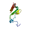
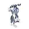
 Structure visualization
 Downloads & links
 Other downloads
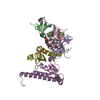


 PDBj
PDBj


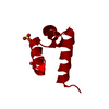
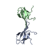
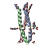
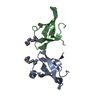
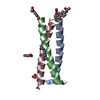
 Assembly
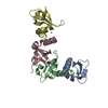
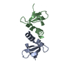
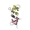


 Num. of mol.: 6 / Source method: obtained synthetically
 Mass: 18.015 Da / Num. of mol.: 115 / Source method: isolated from a natural source / Formula: H2O
 Sample preparation
 Processing