[English] 日本語
Yorodumi
- PDB-5ob8: X-ray structure of the adduct formed upon reaction of lysozyme wi... -

+
Open data


ID or keywords:

Loading...

-
Basic information

Entry
Database: PDB / ID: 5ob8
Title
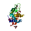
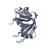
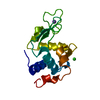
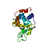
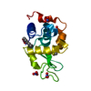
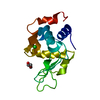
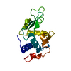
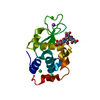
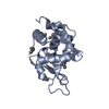
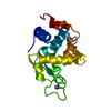
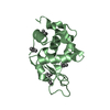
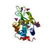
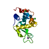
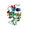
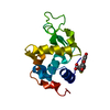
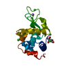
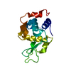
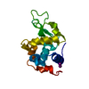
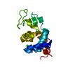
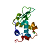
X-ray structure of the adduct formed upon reaction of lysozyme with the compound fac-[RuII(CO)3Cl2(N3-MIM), MIM=methyl-imidazole (crystals grown using NaCl)
ComponentsLysozyme C
KeywordsHYDROLASE / protein-metallodrug interaction / CO releasing molecules
Function / homology
Function and homology information


Antimicrobial peptides / Neutrophil degranulation / beta-N-acetylglucosaminidase activity / cell wall macromolecule catabolic process / lysozyme / lysozyme activity / killing of cells of another organism / defense response to Gram-negative bacterium / defense response to Gram-positive bacterium / defense response to bacterium ...Antimicrobial peptides / Neutrophil degranulation / beta-N-acetylglucosaminidase activity / cell wall macromolecule catabolic process / lysozyme / lysozyme activity / killing of cells of another organism / defense response to Gram-negative bacterium / defense response to Gram-positive bacterium / defense response to bacterium / endoplasmic reticulum / extracellular space / identical protein binding / cytoplasm
Similarity search - Function
Lysozyme - #10 / Glycoside hydrolase, family 22, lysozyme / Glycoside hydrolase family 22 domain / Glycosyl hydrolases family 22 (GH22) domain signature. / Glycoside hydrolase, family 22 / C-type lysozyme/alpha-lactalbumin family / Glycosyl hydrolases family 22 (GH22) domain profile. / Alpha-lactalbumin / lysozyme C / Lysozyme / Lysozyme-like domain superfamily ...Lysozyme - #10 / Glycoside hydrolase, family 22, lysozyme / Glycoside hydrolase family 22 domain / Glycosyl hydrolases family 22 (GH22) domain signature. / Glycoside hydrolase, family 22 / C-type lysozyme/alpha-lactalbumin family / Glycosyl hydrolases family 22 (GH22) domain profile. / Alpha-lactalbumin / lysozyme C / Lysozyme / Lysozyme-like domain superfamily / Orthogonal Bundle / Mainly Alpha
Similarity search - Domain/homology
Chem-9QB / ACETATE ION / Chem-RU2 / Lysozyme C
Similarity search - Component
Biological speciesGallus gallus (chicken)
MethodX-RAY DIFFRACTION / MOLECULAR REPLACEMENT / Resolution: 1.85 Å
AuthorsPontillo, N. / Ferraro, G. / Merlino, A.
CitationJournal: Dalton Trans / Year: 2017
Title: Ru-Based CO releasing molecules with azole ligands: interaction with proteins and the CO release mechanism disclosed by X-ray crystallography.
Authors: Pontillo, N. / Ferraro, G. / Messori, L. / Tamasi, G. / Merlino, A.
History
DepositionJun 26, 2017Deposition site: PDBE / Processing site: PDBE
Revision 1.0Jul 26, 2017Provider: repository / Type: Initial release
Revision 1.1Aug 2, 2017Group: Database references / Category: citation
Item: _citation.country / _citation.journal_volume ..._citation.country / _citation.journal_volume / _citation.page_first / _citation.page_last
Revision 1.2Jan 17, 2024Group: Advisory / Data collection ...Advisory / Data collection / Database references / Derived calculations / Refinement description
Category: chem_comp_atom / chem_comp_bond ...chem_comp_atom / chem_comp_bond / database_2 / pdbx_initial_refinement_model / pdbx_struct_special_symmetry / pdbx_unobs_or_zero_occ_atoms
Item: _database_2.pdbx_DOI / _database_2.pdbx_database_accession

-
Structure visualization

Structure viewerMolecule:
MolmilJmol/JSmol

Downloads & links

-
Assembly

Deposited unit
A: Lysozyme C
hetero molecules


Theoretical massNumber of molelcules
Total (without water)15,0198
Polymers14,3311
Non-polymers6887
Water2,450136
1


  • Idetical with deposited unit
  • defined by software
  • Evidence: homology
TypeNameSymmetry operationNumber
identity operation1_555x,y,z1
Buried area790 Å2
ΔGint-31 kcal/mol
Surface area6570 Å2
MethodPISA
Unit cell
Length a, b, c (Å)78.573, 78.573, 36.664
Angle α, β, γ (deg.)90.00, 90.00, 90.00
Int Tables number96
Space group name H-MP43212
Components on special symmetry positions
IDModelComponents
11A-342-

HOH

21A-348-

HOH

31A-433-

HOH

-
Components

-
Protein , 1 types, 1 molecules A

#1: Protein Lysozyme C / 1 / 4-beta-N-acetylmuramidase C / Allergen Gal d IV


Mass: 14331.160 Da / Num. of mol.: 1 / Source method: isolated from a natural source / Source: (natural) Gallus gallus (chicken) / References: UniProt: P00698, lysozyme

-
Non-polymers , 6 types, 143 molecules

#2: Chemical ChemComp-ACT / ACETATE ION / Acetate


Mass: 59.044 Da / Num. of mol.: 2 / Source method: obtained synthetically / Formula: C2H3O2
#3: Chemical ChemComp-NA / SODIUM ION


Mass: 22.990 Da / Num. of mol.: 1 / Source method: obtained synthetically / Formula: Na
#4: Chemical ChemComp-CL / CHLORIDE ION / Chloride


Mass: 35.453 Da / Num. of mol.: 2 / Source method: obtained synthetically / Formula: Cl
#5: Chemical ChemComp-9QB / chloranyl-bis(oxidaniumyl)-tris(oxidaniumylidynemethyl)ruthenium


Mass: 256.584 Da / Num. of mol.: 1 / Source method: obtained synthetically / Formula: C3H4ClO5Ru
#6: Chemical ChemComp-RU2 / pentakis(oxidaniumyl)-(oxidaniumylidynemethyl)ruthenium


Mass: 219.156 Da / Num. of mol.: 1 / Source method: obtained synthetically / Formula: CH10O6Ru
#7: Water ChemComp-HOH / water / Water


Mass: 18.015 Da / Num. of mol.: 136 / Source method: isolated from a natural source / Formula: H2O

-
Experimental details

-
Experiment

ExperimentMethod: X-RAY DIFFRACTION / Number of used crystals: 1

-
Sample preparation

CrystalDensity Matthews: 1.97 Å3/Da / Density % sol: 37.7 %
Crystal growTemperature: 298 K / Method: vapor diffusion, hanging drop / Details: 1.1M NaCl, 0.1 M sodium acetate pH 4.0

-
Data collection

DiffractionMean temperature: 100 K
Diffraction sourceSource: ROTATING ANODE / Type: RIGAKU MICROMAX-007 HF / Wavelength: 1.5418 Å
DetectorType: RIGAKU SATURN 944 / Detector: CCD / Date: Apr 7, 2014 / Details: mirrors
RadiationProtocol: SINGLE WAVELENGTH / Monochromatic (M) / Laue (L): M / Scattering type: x-ray
Radiation wavelengthWavelength: 1.5418 Å / Relative weight: 1
ReflectionResolution: 1.85→55.56 Å / Num. obs: 9488 / % possible obs: 92.3 % / Redundancy: 4.9 % / Rmerge(I) obs: 0.048 / Net I/σ(I): 36.8
Reflection shellResolution: 1.85→1.88 Å / Rmerge(I) obs: 0.603 / Mean I/σ(I) obs: 2.3

-
Processing

Software
NameVersionClassification
REFMAC5.8.0158refinement
HKL-2000data reduction
HKL-2000data scaling
PHASERphasing
RefinementMethod to determine structure: MOLECULAR REPLACEMENT
Starting model: 193L
Resolution: 1.85→55.56 Å / Cor.coef. Fo:Fc: 0.972 / Cor.coef. Fo:Fc free: 0.937 / SU B: 3.78 / SU ML: 0.111 / Cross valid method: THROUGHOUT / ESU R: 0.165 / ESU R Free: 0.162 / Details: HYDROGENS HAVE BEEN ADDED IN THE RIDING POSITIONS
RfactorNum. reflection% reflectionSelection details
Rfree0.2298 457 4.8 %RANDOM
Rwork0.15955 ---
obs0.163 8981 92.08 %-
Solvent computationIon probe radii: 0.8 Å / Shrinkage radii: 0.8 Å / VDW probe radii: 1.2 Å
Displacement parametersBiso mean: 33.256 Å2
Baniso -1Baniso -2Baniso -3
1-0.01 Å2-0 Å2-0 Å2
2--0.01 Å2-0 Å2
3----0.03 Å2
Refinement stepCycle: 1 / Resolution: 1.85→55.56 Å
ProteinNucleic acidLigandSolventTotal
Num. atoms1000 0 26 136 1162
Refine LS restraints
Refine-IDTypeDev idealDev ideal targetNumber
X-RAY DIFFRACTIONr_bond_refined_d0.0270.021086
X-RAY DIFFRACTIONr_bond_other_d0.0020.02957
X-RAY DIFFRACTIONr_angle_refined_deg1.8381.9521476
X-RAY DIFFRACTIONr_angle_other_deg1.10732212
X-RAY DIFFRACTIONr_dihedral_angle_1_deg7.065137
X-RAY DIFFRACTIONr_dihedral_angle_2_deg37.27123.01953
X-RAY DIFFRACTIONr_dihedral_angle_3_deg14.36215179
X-RAY DIFFRACTIONr_dihedral_angle_4_deg20.1791512
X-RAY DIFFRACTIONr_chiral_restr0.1230.2149
X-RAY DIFFRACTIONr_gen_planes_refined0.0090.021238
X-RAY DIFFRACTIONr_gen_planes_other0.0020.02246
X-RAY DIFFRACTIONr_nbd_refined
X-RAY DIFFRACTIONr_nbd_other
X-RAY DIFFRACTIONr_nbtor_refined
X-RAY DIFFRACTIONr_nbtor_other
X-RAY DIFFRACTIONr_xyhbond_nbd_refined
X-RAY DIFFRACTIONr_xyhbond_nbd_other
X-RAY DIFFRACTIONr_metal_ion_refined
X-RAY DIFFRACTIONr_metal_ion_other
X-RAY DIFFRACTIONr_symmetry_vdw_refined
X-RAY DIFFRACTIONr_symmetry_vdw_other
X-RAY DIFFRACTIONr_symmetry_hbond_refined
X-RAY DIFFRACTIONr_symmetry_hbond_other
X-RAY DIFFRACTIONr_symmetry_metal_ion_refined
X-RAY DIFFRACTIONr_symmetry_metal_ion_other
X-RAY DIFFRACTIONr_mcbond_it2.8123.1535
X-RAY DIFFRACTIONr_mcbond_other2.7653.087532
X-RAY DIFFRACTIONr_mcangle_it3.5374.624669
X-RAY DIFFRACTIONr_mcangle_other3.5354.629670
X-RAY DIFFRACTIONr_scbond_it3.5733.462551
X-RAY DIFFRACTIONr_scbond_other3.4453.439539
X-RAY DIFFRACTIONr_scangle_it
X-RAY DIFFRACTIONr_scangle_other5.0735.015785
X-RAY DIFFRACTIONr_long_range_B_refined6.77437.7671343
X-RAY DIFFRACTIONr_long_range_B_other6.77637.7811344
X-RAY DIFFRACTIONr_rigid_bond_restr
X-RAY DIFFRACTIONr_sphericity_free
X-RAY DIFFRACTIONr_sphericity_bonded
LS refinement shellResolution: 1.85→1.898 Å / Total num. of bins used: 20
RfactorNum. reflection% reflection
Rfree0.366 31 -
Rwork0.256 577 -
obs--85.03 %

+
About Yorodumi

-
News

-
Feb 9, 2022. New format data for meta-information of EMDB entries

New format data for meta-information of EMDB entries

  • Version 3 of the EMDB header file is now the official format.
  • The previous official version 1.9 will be removed from the archive.

Related info.:EMDB header

External links:wwPDB to switch to version 3 of the EMDB data model

-
Aug 12, 2020. Covid-19 info

Covid-19 info

URL: https://pdbj.org/emnavi/covid19.php

New page: Covid-19 featured information page in EM Navigator.

Related info.:Covid-19 info / Mar 5, 2020. Novel coronavirus structure data

+
Mar 5, 2020. Novel coronavirus structure data

Novel coronavirus structure data

Related info.:Yorodumi Speices / Aug 12, 2020. Covid-19 info

External links:COVID-19 featured content - PDBj / Molecule of the Month (242):Coronavirus Proteases

+
Jan 31, 2019. EMDB accession codes are about to change! (news from PDBe EMDB page)

EMDB accession codes are about to change! (news from PDBe EMDB page)

  • The allocation of 4 digits for EMDB accession codes will soon come to an end. Whilst these codes will remain in use, new EMDB accession codes will include an additional digit and will expand incrementally as the available range of codes is exhausted. The current 4-digit format prefixed with “EMD-” (i.e. EMD-XXXX) will advance to a 5-digit format (i.e. EMD-XXXXX), and so on. It is currently estimated that the 4-digit codes will be depleted around Spring 2019, at which point the 5-digit format will come into force.
  • The EM Navigator/Yorodumi systems omit the EMD- prefix.

Related info.:Q: What is EMD? / ID/Accession-code notation in Yorodumi/EM Navigator

External links:EMDB Accession Codes are Changing Soon! / Contact to PDBj

+
Jul 12, 2017. Major update of PDB

Major update of PDB

  • wwPDB released updated PDB data conforming to the new PDBx/mmCIF dictionary.
  • This is a major update changing the version number from 4 to 5, and with Remediation, in which all the entries are updated.
  • In this update, many items about electron microscopy experimental information are reorganized (e.g. em_software).
  • Now, EM Navigator and Yorodumi are based on the updated data.

External links:wwPDB Remediation / Enriched Model Files Conforming to OneDep Data Standards Now Available in the PDB FTP Archive

-
Yorodumi

Thousand views of thousand structures

  • Yorodumi is a browser for structure data from EMDB, PDB, SASBDB, etc.
  • This page is also the successor to EM Navigator detail page, and also detail information page/front-end page for Omokage search.
  • The word "yorodu" (or yorozu) is an old Japanese word meaning "ten thousand". "mi" (miru) is to see.

Related info.:EMDB / PDB / SASBDB / Comparison of 3 databanks / Yorodumi Search / Aug 31, 2016. New EM Navigator & Yorodumi / Yorodumi Papers / Jmol/JSmol / Function and homology information / Changes in new EM Navigator and Yorodumi

Read more