| Entry | Database: PDB / ID: 5iel
|
|---|
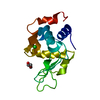
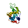
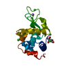
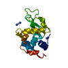
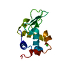
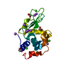
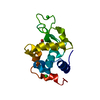
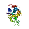
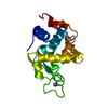
| Title | Structure of Lysozyme labeled with fluorescein isothiocyanate (FITC) at 1.15 Angstroms resolution |
|---|
 Components Components | Lysozyme C |
|---|
 Keywords Keywords | HYDROLASE / glycoside hydrolase muramidase fluorophore |
|---|
| Function / homology |  Function and homology information Function and homology information
Lactose synthesis / Antimicrobial peptides / Neutrophil degranulation / beta-N-acetylglucosaminidase activity / cell wall macromolecule catabolic process / lysozyme / lysozyme activity / killing of cells of another organism / defense response to Gram-negative bacterium / defense response to bacterium ...Lactose synthesis / Antimicrobial peptides / Neutrophil degranulation / beta-N-acetylglucosaminidase activity / cell wall macromolecule catabolic process / lysozyme / lysozyme activity / killing of cells of another organism / defense response to Gram-negative bacterium / defense response to bacterium / defense response to Gram-positive bacterium / endoplasmic reticulum / : / identical protein binding / cytoplasmSimilarity search - Function Lysozyme - #10 / Glycoside hydrolase, family 22, lysozyme / Glycoside hydrolase family 22 domain / Glycosyl hydrolases family 22 (GH22) domain signature. / Glycoside hydrolase, family 22 / C-type lysozyme/alpha-lactalbumin family / Glycosyl hydrolases family 22 (GH22) domain profile. / Alpha-lactalbumin / lysozyme C / Lysozyme / Lysozyme-like domain superfamily ...Lysozyme - #10 / Glycoside hydrolase, family 22, lysozyme / Glycoside hydrolase family 22 domain / Glycosyl hydrolases family 22 (GH22) domain signature. / Glycoside hydrolase, family 22 / C-type lysozyme/alpha-lactalbumin family / Glycosyl hydrolases family 22 (GH22) domain profile. / Alpha-lactalbumin / lysozyme C / Lysozyme / Lysozyme-like domain superfamily / Orthogonal Bundle / Mainly AlphaSimilarity search - Domain/homology |
|---|
| Biological species |   Gallus gallus (chicken) Gallus gallus (chicken) |
|---|
| Method |  X-RAY DIFFRACTION / SYNCHROTRON / MOLECULAR REPLACEMENT / Resolution: 1.15 Å X-RAY DIFFRACTION / SYNCHROTRON / MOLECULAR REPLACEMENT / Resolution: 1.15 Å |
|---|
 Authors Authors | Kachalova, G.S. / Vlaskina, A.V. / Popov, A.P. / Simanovskaia, A.A. / Krukova, M.V. / Lipkin, A.V. |
|---|
| Funding support |  Russian Federation, 1items Russian Federation, 1items | Organization | Grant number | Country |
|---|
| Russian Fund of Fundamental Reasearch | 13-04-12084 | Russian Federation |
|
|---|
 Citation Citation | Journal: To Be Published
Title: Structure of Lysozyme labeled with fluorescein isothiocyanate (FITC) at 1.15 Angstroms resolution
Authors: Kachalova, G.S. / Vlaskina, A.V. / Popov, A.P. / Simanovskaia, A.A. / Krukova, M.V. / Lipkin, A.V. |
|---|
| History | | Deposition | Feb 25, 2016 | Deposition site: RCSB / Processing site: PDBE |
|---|
| Revision 1.0 | Mar 8, 2017 | Provider: repository / Type: Initial release |
|---|
| Revision 1.1 | Jan 10, 2024 | Group: Data collection / Database references / Refinement description
Category: chem_comp_atom / chem_comp_bond ...chem_comp_atom / chem_comp_bond / database_2 / pdbx_initial_refinement_model
Item: _database_2.pdbx_DOI / _database_2.pdbx_database_accession |
|---|
| Revision 1.2 | Oct 23, 2024 | Group: Structure summary / Category: pdbx_entry_details / pdbx_modification_feature |
|---|
|
|---|
 Movie
Movie Controller

 Yorodumi
 Open data
 Basic information
 Structure visualization
 Downloads & links
 Other downloads
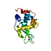

 PDBj
PDBj


 Assembly
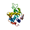
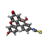
 Mass: 391.397 Da / Num. of mol.: 1 / Source method: isolated from a natural source / Formula: C21H13NO5S
 Mass: 18.015 Da / Num. of mol.: 174 / Source method: isolated from a natural source / Formula: H2O
 Sample preparation
 / Beamline: ID14-1 / Wavelength: 0.97241 Å
 Processing