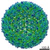
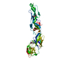
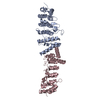
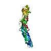
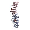
Entry Database : PDB / ID : 5mv1Title Crystal structure of the E protein of the Japanese encephalitis virulent virus E protein Keywords / Function / homology Function Domain/homology Component
/ / / / / / / / / / / / / / / / / / / / / / / / / / / / / / / / / / / / / / / / / / / / / / / / / / / / / / / / / / / / / / / / / / / / / / / / / / / / / / / / / / / / / / / / / / / / / / / / / / / / / / / / / / / / / / / / / / / / / / / / / / / / / / / Biological species Method / / / Resolution : 2.25 Å Authors Liu, X. / Zhao, X. / Na, R. / Li, L. / Warkentin, E. / Witt, J. / Lu, X. / Wei, Y. / Peng, G. / Li, Y. / Wang, J. Journal : Protein Cell / Year : 2019Title : The structure differences of Japanese encephalitis virus SA14 and SA14-14-2 E proteins elucidate the virulence attenuation mechanism.Authors : Liu, X. / Zhao, X. / Na, R. / Li, L. / Warkentin, E. / Witt, J. / Lu, X. / Yu, Y. / Wei, Y. / Peng, G. / Li, Y. / Wang, J. History Deposition Jan 14, 2017 Deposition site / Processing site Revision 1.0 May 23, 2018 Provider / Type Revision 1.1 Feb 27, 2019 Group / Database references / Category / pdbx_database_procItem _citation.journal_volume / _citation.page_first ... _citation.journal_volume / _citation.page_first / _citation.page_last / _citation.year Revision 1.2 Jan 17, 2024 Group / Database references / Refinement descriptionCategory chem_comp_atom / chem_comp_bond ... chem_comp_atom / chem_comp_bond / database_2 / pdbx_initial_refinement_model Item / _database_2.pdbx_database_accession
Show all Show less
 Movie
Movie Controller

 Yorodumi
 Open data
 Basic information
 Components
 Keywords
 Function and homology information
 Japanese encephalitis virus
 X-RAY DIFFRACTION /
  Authors
 Citation
 Structure visualization
 Downloads & links
 Other downloads
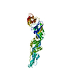


 PDBj
PDBj


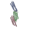
 Assembly

 Mass: 18.015 Da / Num. of mol.: 115 / Source method: isolated from a natural source / Formula: H2O
 Sample preparation
 / Beamline: BL18U1 / Wavelength: 0.97776 Å
 Processing