Protocol: SINGLE WAVELENGTH / Monochromatic (M) / Laue (L): M / Scattering type: x-ray
Radiation wavelength
ID
Wavelength (Å)
Relative weight
1
1
1
2
0.9792
1
Reflection
Resolution: 1.9→55.33 Å / Num. obs: 13677 / % possible obs: 97.4 % / Redundancy: 3.2 % / Rmerge(I) obs: 0.07 / Net I/σ(I): 14
Reflection shell
Resolution: 1.9→1.93 Å / Redundancy: 3.3 % / Rmerge(I) obs: 0.458 / % possible all: 96.2
-
Processing
Software
Name
Version
Classification
REFMAC
5.8.0103
refinement
HKL-2000
datareduction
HKL-2000
datascaling
PHENIX
phasing
Refinement
Method to determine structure: SAD / Resolution: 1.91→55.33 Å / Cor.coef. Fo:Fc: 0.951 / Cor.coef. Fo:Fc free: 0.922 / SU B: 7.881 / SU ML: 0.124 / Cross valid method: THROUGHOUT / ESU R: 0.178 / ESU R Free: 0.165 / Details: HYDROGENS HAVE BEEN ADDED IN THE RIDING POSITIONS
Rfactor
Num. reflection
% reflection
Selection details
Rfree
0.24615
686
5 %
RANDOM
Rwork
0.19444
-
-
-
obs
0.19698
12989
96.45 %
-
Solvent computation
Ion probe radii: 0.8 Å / Shrinkage radii: 0.8 Å / VDW probe radii: 1.2 Å
 Movie
Movie Controller

 Yorodumi
 Open data
 Basic information
 Components
 Keywords
 Function and homology information
 X-RAY DIFFRACTION /
  Authors
 Japan, 1items
  Citation
 Structure visualization
 Downloads & links
 Other downloads
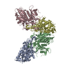


 PDBj
PDBj
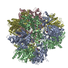
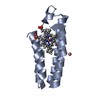
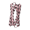
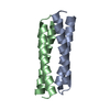
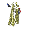
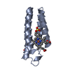
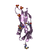
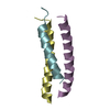
 Assembly


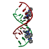
 Mass: 18.015 Da / Num. of mol.: 176 / Source method: isolated from a natural source / Formula: H2O
 Sample preparation
 Processing