 Movie
Movie Controller

[English] 日本語
 Yorodumi
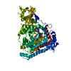
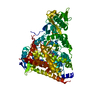
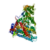
- PDB-5h8u: Crystal structure of mycobacterium tuberculosis wild-type malate ... -
+ Open data

- Basic information
| Entry | Database: PDB / ID: 5h8u | ||||||
|---|---|---|---|---|---|---|---|
| Title | Crystal structure of mycobacterium tuberculosis wild-type malate synthase in complex with product malate | ||||||
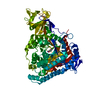
 Components Components | Malate synthase G | ||||||
 Keywords Keywords | TRANSFERASE | ||||||
| Function / homology |  Function and homology information Function and homology informationhost cell extracellular matrix binding / capsule / malate synthase / malate synthase activity / glyoxylate catabolic process / adhesion of symbiont to host / coenzyme A binding / coenzyme A metabolic process / glyoxylate cycle / fibronectin binding ...host cell extracellular matrix binding / capsule / malate synthase / malate synthase activity / glyoxylate catabolic process / adhesion of symbiont to host / coenzyme A binding / coenzyme A metabolic process / glyoxylate cycle / fibronectin binding / tricarboxylic acid cycle / laminin binding / peptidoglycan-based cell wall / magnesium ion binding / cell surface / protein homodimerization activity / extracellular region / plasma membrane / cytoplasm / cytosol Similarity search - Function | ||||||
| Biological species |   Mycobacterium tuberculosis (bacteria) Mycobacterium tuberculosis (bacteria) | ||||||
| Method |  X-RAY DIFFRACTION / SYNCHROTRON / MOLECULAR REPLACEMENT / Resolution: 2.85 Å X-RAY DIFFRACTION / SYNCHROTRON / MOLECULAR REPLACEMENT / Resolution: 2.85 Å | ||||||
 Authors Authors | Krieger, I.V. / Huang, H.-L. / Sacchettini, J.C. | ||||||
| Funding support |  United States, 1items United States, 1items
| ||||||
 Citation Citation | Journal: J. Biol. Chem. / Year: 2016 Title: Mycobacterium tuberculosis Malate Synthase Structures with Fragments Reveal a Portal for Substrate/Product Exchange. Authors: Huang, H.L. / Krieger, I.V. / Parai, M.K. / Gawandi, V.B. / Sacchettini, J.C. #1: Journal: J.Biol.Chem. / Year: 2003Title: Biochemical and structural studies of malate synthase from Mycobacterium tuberculosis. Authors: Smith, C.V. / Huang, C.C. / Miczak, A. / Russell, D.G. / Sacchettini, J.C. / Honer zu Bentrup, K. #2: Journal: Chem.Biol. / Year: 2012Title: Structure-guided discovery of phenyl-diketo acids as potent inhibitors of M. tuberculosis malate synthase. Authors: Krieger, I.V. / Freundlich, J.S. / Gawandi, V.B. / Roberts, J.P. / Gawandi, V.B. / Sun, Q. / Owen, J.L. / Fraile, M.T. / Huss, S.I. / Lavandera, J.L. / Ioerger, T.R. / Sacchettini, J.C. | ||||||
| History |
|
- Structure visualization
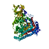
| Structure viewer | Molecule: MolmilJmol/JSmol |
|---|
- Downloads & links
-Download
| PDBx/mmCIF format | 5h8u.cif.gz | 283.6 KB | Display | PDBx/mmCIF format |
|---|---|---|---|---|
| PDB format | pdb5h8u.ent.gz | 228.1 KB | Display | PDB format |
| PDBx/mmJSON format | 5h8u.json.gz | Tree view | PDBx/mmJSON format | |
| Others |  Other downloads Other downloads |
-Validation report
| Arichive directory | https://data.pdbj.org/pub/pdb/validation_reports/h8/5h8uftp://data.pdbj.org/pub/pdb/validation_reports/h8/5h8u | HTTPS FTP |
|---|
-Related structure data
| Related structure data |  5c7vC 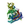 5c9rC  5c9uC  5c9wC  5c9xC  5cahC  5cakC  5cbbC  5cbiC  5cbjC  5cczC  5cewC  5cjmC  5cjnC  5drcC  5driC  5dx7C 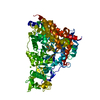 5e9xC  5ecvC  5h8mC 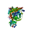 5h8pC  5t8gC  1n8iS C: citing same article ( S: Starting model for refinement |
|---|---|
| Similar structure data |


-Links
 PDBj
PDBj
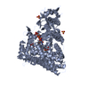
- Assembly
| Deposited unit | 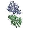
| ||||||||
|---|---|---|---|---|---|---|---|---|---|
| 1 | 
| ||||||||
| 2 | 
| ||||||||
| Unit cell |
|
-Components
| #1: Protein | Mass: 80488.797 Da / Num. of mol.: 2 Source method: isolated from a genetically manipulated source Details: Regions containing residues 1-2, 304-308, and 728-741 are disordered and excluded from the final refined model. Source: (gene. exp.) Mycobacterium tuberculosis (strain ATCC 25618 / H37Rv) (bacteria)Strain: ATCC 25618 / H37Rv / Gene: glcB, Rv1837c, MTCY1A11.06 / Production host: #2: Chemical |   Mass: 24.305 Da / Num. of mol.: 3 / Source method: obtained synthetically / Formula: Mg Mass: 24.305 Da / Num. of mol.: 3 / Source method: obtained synthetically / Formula: Mg#3: Chemical | ChemComp-LMR / ( |   Mass: 134.087 Da / Num. of mol.: 1 / Source method: obtained synthetically / Formula: C4H6O5 Mass: 134.087 Da / Num. of mol.: 1 / Source method: obtained synthetically / Formula: C4H6O5#4: Chemical | ChemComp-GLV / | 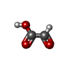  Mass: 74.035 Da / Num. of mol.: 1 / Source method: obtained synthetically / Formula: C2H2O3 Mass: 74.035 Da / Num. of mol.: 1 / Source method: obtained synthetically / Formula: C2H2O3#5: Water | ChemComp-HOH / |  Mass: 18.015 Da / Num. of mol.: 60 / Source method: isolated from a natural source / Formula: H2O Mass: 18.015 Da / Num. of mol.: 60 / Source method: isolated from a natural source / Formula: H2O |
|---|
-Experimental details
-Experiment
| Experiment | Method: X-RAY DIFFRACTION / Number of used crystals: 1 |
|---|
- Sample preparation
| Crystal | Density Matthews: 2.63 Å3/Da / Density % sol: 53.23 % |
|---|---|
| Crystal grow | Temperature: 290 K / Method: vapor diffusion, hanging drop / pH: 7.5 / Details: PEG 3350, magnesium chloride, tris / PH range: 7.0-8.5 / Temp details: Varies between 289-291 |
-Data collection
| Diffraction | Mean temperature: 100 K |
|---|---|
| Diffraction source | Source: SYNCHROTRON / Site: APS / Beamline: 19-ID / Wavelength: 0.98 Å |
| Detector | Type: ADSC QUANTUM 315 / Detector: CCD / Date: Nov 18, 2013 |
| Radiation | Monochromator: Si(111) / Protocol: SINGLE WAVELENGTH / Monochromatic (M) / Laue (L): M / Scattering type: x-ray |
| Radiation wavelength | Wavelength: 0.98 Å / Relative weight: 1 |
| Reflection | Resolution: 2.85→37.362 Å / Num. obs: 40032 / % possible obs: 98.2 % / Redundancy: 14.4 % / Rmerge(I) obs: 0.105 / Rsym value: 0.0516 / Net I/σ(I): 14.96 |
| Reflection shell | Resolution: 2.85→2.9 Å / Redundancy: 13.6 % / Rmerge(I) obs: 0.997 / Mean I/σ(I) obs: 4.58 / % possible all: 100 |
- Processing
| Software |
| ||||||||||||||||||||||||||||||||||||||||||||||||||||||||||||||||||||||||||||||||||||||||||||||||||||||||||||||||
|---|---|---|---|---|---|---|---|---|---|---|---|---|---|---|---|---|---|---|---|---|---|---|---|---|---|---|---|---|---|---|---|---|---|---|---|---|---|---|---|---|---|---|---|---|---|---|---|---|---|---|---|---|---|---|---|---|---|---|---|---|---|---|---|---|---|---|---|---|---|---|---|---|---|---|---|---|---|---|---|---|---|---|---|---|---|---|---|---|---|---|---|---|---|---|---|---|---|---|---|---|---|---|---|---|---|---|---|---|---|---|---|---|---|
| Refinement | Method to determine structure: MOLECULAR REPLACEMENT Starting model: 1N8I Resolution: 2.85→37.362 Å / SU ML: 0.42 / Cross valid method: FREE R-VALUE / σ(F): 1.34 / Phase error: 32.05 / Stereochemistry target values: ML
| ||||||||||||||||||||||||||||||||||||||||||||||||||||||||||||||||||||||||||||||||||||||||||||||||||||||||||||||||
| Solvent computation | Shrinkage radii: 0.9 Å / VDW probe radii: 1.11 Å / Solvent model: FLAT BULK SOLVENT MODEL | ||||||||||||||||||||||||||||||||||||||||||||||||||||||||||||||||||||||||||||||||||||||||||||||||||||||||||||||||
| Refinement step | Cycle: LAST / Resolution: 2.85→37.362 Å
| ||||||||||||||||||||||||||||||||||||||||||||||||||||||||||||||||||||||||||||||||||||||||||||||||||||||||||||||||
| Refine LS restraints |
| ||||||||||||||||||||||||||||||||||||||||||||||||||||||||||||||||||||||||||||||||||||||||||||||||||||||||||||||||
| LS refinement shell |
|
