 Movie
Movie Controller

[English] 日本語
 Yorodumi
- PDB-5h6s: Crystal structure of Hydrazidase S179A mutant complexed with a su... -
+ Open data

- Basic information
| Entry | Database: PDB / ID: 5h6s | ||||||
|---|---|---|---|---|---|---|---|
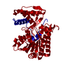
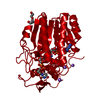
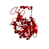
| Title | Crystal structure of Hydrazidase S179A mutant complexed with a substrate | ||||||
 Components Components | Amidase | ||||||
 Keywords Keywords | HYDROLASE / amidase | ||||||
| Function / homology | Amidase / Amidase, conserved site / Amidases signature. / Amidase signature domain / Amidase signature (AS) superfamily / Amidase / amidase activity / 4-oxidanylbenzohydrazide / Amidase Function and homology information Function and homology information | ||||||
| Biological species |  Microbacterium sp. HM58-2 (bacteria) Microbacterium sp. HM58-2 (bacteria) | ||||||
| Method |  X-RAY DIFFRACTION / SYNCHROTRON / MOLECULAR REPLACEMENT / Resolution: 1.8 Å X-RAY DIFFRACTION / SYNCHROTRON / MOLECULAR REPLACEMENT / Resolution: 1.8 Å | ||||||
 Authors Authors | Akiyama, T. / Ishii, M. / Takuwa, A. / Oinuma, K. / Sasaki, Y. / Takaya, N. / Yajima, S. | ||||||
 Citation Citation | Journal: Biochem. Biophys. Res. Commun. / Year: 2017 Title: Structural basis of the substrate recognition of hydrazidase isolated from Microbacterium sp. strain HM58-2, which catalyzes acylhydrazide compounds as its sole carbon source Authors: Akiyama, T. / Ishii, M. / Takuwa, A. / Oinuma, K.I. / Sasaki, Y. / Takaya, N. / Yajima, S. | ||||||
| History |
|
- Structure visualization
| Structure viewer | Molecule: MolmilJmol/JSmol |
|---|
- Downloads & links
-Download
| PDBx/mmCIF format | 5h6s.cif.gz | 357.5 KB | Display | PDBx/mmCIF format |
|---|---|---|---|---|
| PDB format | pdb5h6s.ent.gz | 288.9 KB | Display | PDB format |
| PDBx/mmJSON format | 5h6s.json.gz | Tree view | PDBx/mmJSON format | |
| Others |  Other downloads Other downloads |
-Validation report
| Summary document | 5h6s_validation.pdf.gz | 478.4 KB | Display | wwPDB validaton report |
|---|---|---|---|---|
| Full document | 5h6s_full_validation.pdf.gz | 484.1 KB | Display | |
| Data in XML | 5h6s_validation.xml.gz | 66.8 KB | Display | |
| Data in CIF | 5h6s_validation.cif.gz | 96.2 KB | Display | |
| Arichive directory | https://data.pdbj.org/pub/pdb/validation_reports/h6/5h6sftp://data.pdbj.org/pub/pdb/validation_reports/h6/5h6s | HTTPS FTP |
-Related structure data
| Related structure data | 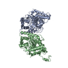 5h6tSC S: Starting model for refinement C: citing same article ( |
|---|---|
| Similar structure data |


-Links
 PDBj
PDBj- Assembly
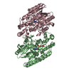
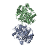
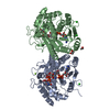
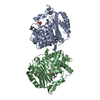
| Deposited unit | 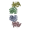
| ||||||||
|---|---|---|---|---|---|---|---|---|---|
| 1 | 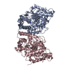
| ||||||||
| 2 | 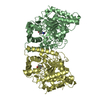
| ||||||||
| Unit cell |
|
-Components
| #1: Protein | Mass: 51565.133 Da / Num. of mol.: 4 Source method: isolated from a genetically manipulated source Source: (gene. exp.) Microbacterium sp. HM58-2 (bacteria) / Strain: HM58-2 / Gene: MHM582_3487 / Production host: #2: Chemical | ChemComp-HDH / 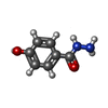  Mass: 152.151 Da / Num. of mol.: 4 / Source method: obtained synthetically / Formula: C7H8N2O2 Mass: 152.151 Da / Num. of mol.: 4 / Source method: obtained synthetically / Formula: C7H8N2O2#3: Water | ChemComp-HOH / |  Mass: 18.015 Da / Num. of mol.: 727 / Source method: isolated from a natural source / Formula: H2O Mass: 18.015 Da / Num. of mol.: 727 / Source method: isolated from a natural source / Formula: H2O |
|---|
-Experimental details
-Experiment
| Experiment | Method: X-RAY DIFFRACTION / Number of used crystals: 1 |
|---|
- Sample preparation
| Crystal | Density Matthews: 2.66 Å3/Da / Density % sol: 53.69 % |
|---|---|
| Crystal grow | Temperature: 277 K / Method: vapor diffusion, hanging drop / Details: 0.1M imidazole, 0.17 M LiSO4, 2M (NH4)2SO4 |
-Data collection
| Diffraction | Mean temperature: 95 K |
|---|---|
| Diffraction source | Source: SYNCHROTRON / Site: Photon Factory  / Beamline: BL-17A / Wavelength: 0.98 Å / Beamline: BL-17A / Wavelength: 0.98 Å |
| Detector | Type: ADSC QUANTUM 270 / Detector: CCD / Date: Jun 22, 2013 |
| Radiation | Protocol: SINGLE WAVELENGTH / Monochromatic (M) / Laue (L): M / Scattering type: x-ray |
| Radiation wavelength | Wavelength: 0.98 Å / Relative weight: 1 |
| Reflection | Resolution: 1.8→50 Å / Num. obs: 196526 / % possible obs: 97.9 % / Redundancy: 3.8 % / Rmerge(I) obs: 0.065 / Net I/σ(I): 29.9 |
| Reflection shell | Resolution: 1.8→1.82 Å / Redundancy: 3.7 % / Rmerge(I) obs: 0.386 / Mean I/σ(I) obs: 5.2 / % possible all: 96.3 |
- Processing
| Software |
| ||||||||||||||||||||||||||||||||||||||||||||||||||||||||||||||||||||||||||||||||||||||||||||||||||||||||||||||||||||||||||||||||||||||||||||||||||||||||||||||||||||||||||||||||||||||
|---|---|---|---|---|---|---|---|---|---|---|---|---|---|---|---|---|---|---|---|---|---|---|---|---|---|---|---|---|---|---|---|---|---|---|---|---|---|---|---|---|---|---|---|---|---|---|---|---|---|---|---|---|---|---|---|---|---|---|---|---|---|---|---|---|---|---|---|---|---|---|---|---|---|---|---|---|---|---|---|---|---|---|---|---|---|---|---|---|---|---|---|---|---|---|---|---|---|---|---|---|---|---|---|---|---|---|---|---|---|---|---|---|---|---|---|---|---|---|---|---|---|---|---|---|---|---|---|---|---|---|---|---|---|---|---|---|---|---|---|---|---|---|---|---|---|---|---|---|---|---|---|---|---|---|---|---|---|---|---|---|---|---|---|---|---|---|---|---|---|---|---|---|---|---|---|---|---|---|---|---|---|---|---|
| Refinement | Method to determine structure: MOLECULAR REPLACEMENT Starting model: 5H6T Resolution: 1.8→40.6 Å / Cor.coef. Fo:Fc: 0.957 / Cor.coef. Fo:Fc free: 0.948 / SU B: 2.058 / SU ML: 0.065 / Cross valid method: THROUGHOUT / ESU R: 0.107 / ESU R Free: 0.098 / Details: HYDROGENS HAVE BEEN ADDED IN THE RIDING POSITIONS
| ||||||||||||||||||||||||||||||||||||||||||||||||||||||||||||||||||||||||||||||||||||||||||||||||||||||||||||||||||||||||||||||||||||||||||||||||||||||||||||||||||||||||||||||||||||||
| Solvent computation | Ion probe radii: 0.8 Å / Shrinkage radii: 0.8 Å / VDW probe radii: 1.2 Å | ||||||||||||||||||||||||||||||||||||||||||||||||||||||||||||||||||||||||||||||||||||||||||||||||||||||||||||||||||||||||||||||||||||||||||||||||||||||||||||||||||||||||||||||||||||||
| Displacement parameters | Biso mean: 18.018 Å2
| ||||||||||||||||||||||||||||||||||||||||||||||||||||||||||||||||||||||||||||||||||||||||||||||||||||||||||||||||||||||||||||||||||||||||||||||||||||||||||||||||||||||||||||||||||||||
| Refinement step | Cycle: 1 / Resolution: 1.8→40.6 Å
| ||||||||||||||||||||||||||||||||||||||||||||||||||||||||||||||||||||||||||||||||||||||||||||||||||||||||||||||||||||||||||||||||||||||||||||||||||||||||||||||||||||||||||||||||||||||
| Refine LS restraints |
|