Monochromator: DIAMOND (111), GE(220) / Protocol: SINGLE WAVELENGTH / Monochromatic (M) / Laue (L): M / Scattering type: x-ray
Radiation wavelength
Wavelength: 0.933 Å / Relative weight: 1
Reflection
Resolution: 2.3→87.7 Å / Num. obs: 25160 / % possible obs: 97.8 % / Observed criterion σ(I): 3 / Redundancy: 16.3 % / Rmerge(I) obs: 0.07 / Net I/σ(I): 7.8
Reflection shell
Resolution: 2.3→2.42 Å / Redundancy: 11.4 % / Rmerge(I) obs: 0.37 / Mean I/σ(I) obs: 2.1 / % possible all: 97.8
-
Processing
Software
Name
Version
Classification
REFMAC
5.2.0007
refinement
MOSFLM
datareduction
SCALA
datascaling
SOLVE
phasing
Refinement
Method to determine structure: MAD / Resolution: 2.3→87.7 Å / Cor.coef. Fo:Fc: 0.955 / Cor.coef. Fo:Fc free: 0.937 / SU B: 9.846 / SU ML: 0.125 / TLS residual ADP flag: LIKELY RESIDUAL / Cross valid method: THROUGHOUT / ESU R: 0.209 / ESU R Free: 0.187 / Stereochemistry target values: MAXIMUM LIKELIHOOD Details: HYDROGENS HAVE BEEN ADDED IN THE RIDING POSITIONS. A LOOP CONTAINING RESIDUES V604-D615 IS MISSING IN THE STRUCTURE. THE C-TERMINAL RESIDUES K658-S660 ARE MISSING IN THE STRUCTURE
Rfactor
Num. reflection
% reflection
Selection details
Rfree
0.23
1275
5.1 %
RANDOM
Rwork
0.186
-
-
-
obs
0.188
23801
96.8 %
-
Solvent computation
Ion probe radii: 0.8 Å / Shrinkage radii: 0.8 Å / VDW probe radii: 1.2 Å / Solvent model: BABINET MODEL WITH MASK
 Movie
Movie Controller


 Open data
 Basic information
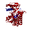
 Components
 Keywords
 Function and homology information
 X-RAY DIFFRACTION /
  Authors
 Citation
 Structure visualization
 Downloads & links
 Other downloads

 PDBj
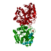
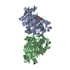
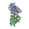
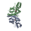
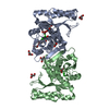
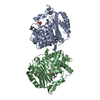
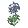
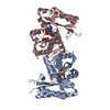
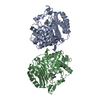
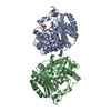
PDBj Assembly
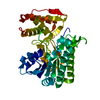
 Mass: 18.015 Da / Num. of mol.: 94 / Source method: isolated from a natural source / Formula: H2O
 Sample preparation
 / Beamline: ID14-2 / Wavelength: 0.933
  Processing