 Movie
Movie Controller

[English] 日本語
 Yorodumi
- PDB-5h3b: Crystal Structure of SeMet-BioG from Haemophilus influenzae at 1.... -
+ Open data

- Basic information
| Entry | Database: PDB / ID: 5h3b | ||||||
|---|---|---|---|---|---|---|---|
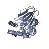
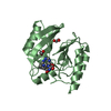
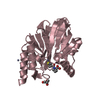
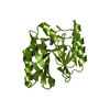
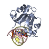
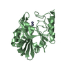
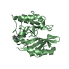
| Title | Crystal Structure of SeMet-BioG from Haemophilus influenzae at 1.49 Angstroms resolution | ||||||
 Components Components | Uncharacterized protein HI_1552 | ||||||
 Keywords Keywords | HYDROLASE / alpha/beta-hydrolase fold / pimeloyl-ACP methyl esterase / biotin biosynthesis | ||||||
| Function / homology | Pimeloyl-ACP methyl esterase BioG / Pimeloyl-ACP methyl esterase BioG / ISOPROPYL ALCOHOL / Uncharacterized protein HI_1552 Function and homology information Function and homology information | ||||||
| Biological species |  Haemophilus influenzae Rd KW20 (bacteria) Haemophilus influenzae Rd KW20 (bacteria) | ||||||
| Method |  X-RAY DIFFRACTION / SYNCHROTRON / SAD / Resolution: 1.492 Å X-RAY DIFFRACTION / SYNCHROTRON / SAD / Resolution: 1.492 Å | ||||||
 Authors Authors | Shi, J. / Guo, Z. | ||||||
 Citation Citation | Journal: Biochemistry / Year: 2016 Title: An Atypical alpha / beta-Hydrolase Fold Revealed in the Crystal Structure of Pimeloyl-Acyl Carrier Protein Methyl Esterase BioG from Haemophilus influenzae Authors: Shi, J. / Cao, X. / Chen, Y. / Cronan, J.E. / Guo, Z. | ||||||
| History |
|
- Structure visualization
| Structure viewer | Molecule: MolmilJmol/JSmol |
|---|
- Downloads & links
-Download
| PDBx/mmCIF format | 5h3b.cif.gz | 198.3 KB | Display | PDBx/mmCIF format |
|---|---|---|---|---|
| PDB format | pdb5h3b.ent.gz | 156.7 KB | Display | PDB format |
| PDBx/mmJSON format | 5h3b.json.gz | Tree view | PDBx/mmJSON format | |
| Others |  Other downloads Other downloads |
-Validation report
| Arichive directory | https://data.pdbj.org/pub/pdb/validation_reports/h3/5h3bftp://data.pdbj.org/pub/pdb/validation_reports/h3/5h3b | HTTPS FTP |
|---|
-Related structure data


-Links
 PDBj
PDBj
- Assembly
| Deposited unit | 
| ||||||||
|---|---|---|---|---|---|---|---|---|---|
| 1 | 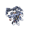
| ||||||||
| 2 | 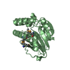
| ||||||||
| Unit cell |
|
-Components
| #1: Protein | Mass: 26443.428 Da / Num. of mol.: 2 Source method: isolated from a genetically manipulated source Source: (gene. exp.) Haemophilus influenzae Rd KW20 (bacteria)Strain: KW20 / Gene: HI_1552 / Production host: #2: Chemical | ChemComp-GOL / |   Mass: 92.094 Da / Num. of mol.: 1 / Source method: obtained synthetically / Formula: C3H8O3 Mass: 92.094 Da / Num. of mol.: 1 / Source method: obtained synthetically / Formula: C3H8O3#3: Chemical | ChemComp-IPA / |   Mass: 60.095 Da / Num. of mol.: 1 / Source method: obtained synthetically / Formula: C3H8O / Comment: alkaloid*YM Mass: 60.095 Da / Num. of mol.: 1 / Source method: obtained synthetically / Formula: C3H8O / Comment: alkaloid*YM#4: Water | ChemComp-HOH / |  Mass: 18.015 Da / Num. of mol.: 470 / Source method: isolated from a natural source / Formula: H2O Mass: 18.015 Da / Num. of mol.: 470 / Source method: isolated from a natural source / Formula: H2OHas protein modification | Y | |
|---|
-Experimental details
-Experiment
| Experiment | Method: X-RAY DIFFRACTION / Number of used crystals: 1 |
|---|
- Sample preparation
| Crystal | Density Matthews: 2.19 Å3/Da / Density % sol: 43.73 % |
|---|---|
| Crystal grow | Temperature: 289 K / Method: vapor diffusion, hanging drop / pH: 5.6 Details: 10%(v/v) iso-propanol, 0.1M citric acid/sodium citrate pH 5.6, 22% PEG 4000 |
-Data collection
| Diffraction | Mean temperature: 100 K | ||||||||||||||||||||||||||||||||||||||||||||||||||||||||||||||||||
|---|---|---|---|---|---|---|---|---|---|---|---|---|---|---|---|---|---|---|---|---|---|---|---|---|---|---|---|---|---|---|---|---|---|---|---|---|---|---|---|---|---|---|---|---|---|---|---|---|---|---|---|---|---|---|---|---|---|---|---|---|---|---|---|---|---|---|---|
| Diffraction source | Source: SYNCHROTRON / Site: SSRF  / Beamline: BL17U / Wavelength: 0.9792 Å / Beamline: BL17U / Wavelength: 0.9792 Å | ||||||||||||||||||||||||||||||||||||||||||||||||||||||||||||||||||
| Detector | Type: ADSC QUANTUM 315r / Detector: CCD / Date: Dec 25, 2015 | ||||||||||||||||||||||||||||||||||||||||||||||||||||||||||||||||||
| Radiation | Protocol: SINGLE WAVELENGTH / Monochromatic (M) / Laue (L): M / Scattering type: x-ray | ||||||||||||||||||||||||||||||||||||||||||||||||||||||||||||||||||
| Radiation wavelength | Wavelength: 0.9792 Å / Relative weight: 1 | ||||||||||||||||||||||||||||||||||||||||||||||||||||||||||||||||||
| Reflection | Resolution: 1.49→32.45 Å / Num. obs: 70407 / % possible obs: 99.6 % / Redundancy: 7.4 % / CC1/2: 0.998 / Rmerge(I) obs: 0.082 / Net I/av σ(I): 19.51 / Net I/σ(I): 10.9 | ||||||||||||||||||||||||||||||||||||||||||||||||||||||||||||||||||
| Reflection shell |
|
- Processing
| Software |
| |||||||||||||||||||||||||||||||||||||||||||||||||||||||||||||||||||||||||||||||||||||||||||||||||||||||||
|---|---|---|---|---|---|---|---|---|---|---|---|---|---|---|---|---|---|---|---|---|---|---|---|---|---|---|---|---|---|---|---|---|---|---|---|---|---|---|---|---|---|---|---|---|---|---|---|---|---|---|---|---|---|---|---|---|---|---|---|---|---|---|---|---|---|---|---|---|---|---|---|---|---|---|---|---|---|---|---|---|---|---|---|---|---|---|---|---|---|---|---|---|---|---|---|---|---|---|---|---|---|---|---|---|---|---|
| Refinement | Method to determine structure: SAD / Resolution: 1.492→32.45 Å / SU ML: 0.15 / Cross valid method: FREE R-VALUE / σ(F): 0 / Phase error: 19.76
| |||||||||||||||||||||||||||||||||||||||||||||||||||||||||||||||||||||||||||||||||||||||||||||||||||||||||
| Solvent computation | Shrinkage radii: 0.9 Å / VDW probe radii: 1.11 Å | |||||||||||||||||||||||||||||||||||||||||||||||||||||||||||||||||||||||||||||||||||||||||||||||||||||||||
| Displacement parameters | Biso max: 65.8 Å2 / Biso mean: 25.32 Å2 / Biso min: 8.42 Å2 | |||||||||||||||||||||||||||||||||||||||||||||||||||||||||||||||||||||||||||||||||||||||||||||||||||||||||
| Refinement step | Cycle: final / Resolution: 1.492→32.45 Å
| |||||||||||||||||||||||||||||||||||||||||||||||||||||||||||||||||||||||||||||||||||||||||||||||||||||||||
| Refine LS restraints |
| |||||||||||||||||||||||||||||||||||||||||||||||||||||||||||||||||||||||||||||||||||||||||||||||||||||||||
| LS refinement shell | Refine-ID: X-RAY DIFFRACTION / Total num. of bins used: 14
|
