 Movie
Movie Controller


+ Open data

- Basic information
| Entry | Database: PDB / ID: 5e9f | ||||||
|---|---|---|---|---|---|---|---|
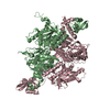
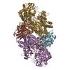
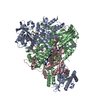
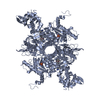
| Title | Structural insights of isocitrate lyases from Magnaporthe oryzae | ||||||
 Components Components | Isocitrate lyase | ||||||
 Keywords Keywords | LYASE / TIM BETA/ALPHA-BARREL / LYASE ACTIVITY | ||||||
| Function / homology |  Function and homology information Function and homology informationglyoxysome / methylisocitrate lyase / isocitrate lyase / methylisocitrate lyase activity / isocitrate lyase activity / glyoxylate cycle / tricarboxylic acid cycle / metal ion binding Similarity search - Function | ||||||
| Biological species |  Magnaporthe oryzae 70-15 (fungus) Magnaporthe oryzae 70-15 (fungus) | ||||||
| Method |  X-RAY DIFFRACTION / SYNCHROTRON / MOLECULAR REPLACEMENT / Resolution: 2.8 Å X-RAY DIFFRACTION / SYNCHROTRON / MOLECULAR REPLACEMENT / Resolution: 2.8 Å | ||||||
 Authors Authors | Park, Y. / Cho, Y. / Lee, Y.-H. / Lee, Y.-W. / Rhee, S. | ||||||
 Citation Citation | Journal: J.Struct.Biol. / Year: 2016 Title: Crystal structure and functional analysis of isocitrate lyases from Magnaporthe oryzae and Fusarium graminearum Authors: Park, Y. / Cho, Y. / Lee, Y.-H. / Lee, Y.-W. / Rhee, S. | ||||||
| History |
|
- Structure visualization
| Structure viewer | Molecule: MolmilJmol/JSmol |
|---|
- Downloads & links
-Download
| PDBx/mmCIF format | 5e9f.cif.gz | 376.1 KB | Display | PDBx/mmCIF format |
|---|---|---|---|---|
| PDB format | pdb5e9f.ent.gz | 303.7 KB | Display | PDB format |
| PDBx/mmJSON format | 5e9f.json.gz | Tree view | PDBx/mmJSON format | |
| Others |  Other downloads Other downloads |
-Validation report
| Arichive directory | https://data.pdbj.org/pub/pdb/validation_reports/e9/5e9fftp://data.pdbj.org/pub/pdb/validation_reports/e9/5e9f | HTTPS FTP |
|---|
-Related structure data
| Related structure data |  5e9gC  5e9hC  1dquS C: citing same article ( S: Starting model for refinement |
|---|---|
| Similar structure data |


-Links
 PDBj
PDBj
- Assembly
| Deposited unit | 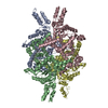
| ||||||||
|---|---|---|---|---|---|---|---|---|---|
| 1 |
| ||||||||
| Unit cell |
|
-Components
| #1: Protein | Mass: 62639.816 Da / Num. of mol.: 4 Source method: isolated from a genetically manipulated source Source: (gene. exp.) Magnaporthe oryzae 70-15 (fungus) / Strain: 70-15 / Gene: ICL1, MGG_04895 / Production host:  References: UniProt: P0CT06, isocitrate lyase, methylisocitrate lyase #2: Chemical | ChemComp-MG /   Mass: 24.305 Da / Num. of mol.: 4 / Source method: obtained synthetically / Formula: Mg Mass: 24.305 Da / Num. of mol.: 4 / Source method: obtained synthetically / Formula: Mg#3: Water | ChemComp-HOH / |  Mass: 18.015 Da / Num. of mol.: 65 / Source method: isolated from a natural source / Formula: H2O Mass: 18.015 Da / Num. of mol.: 65 / Source method: isolated from a natural source / Formula: H2O |
|---|
-Experimental details
-Experiment
| Experiment | Method: X-RAY DIFFRACTION / Number of used crystals: 1 |
|---|
- Sample preparation
| Crystal | Density Matthews: 2.67 Å3/Da / Density % sol: 54.01 % |
|---|---|
| Crystal grow | Temperature: 295 K / Method: vapor diffusion, sitting drop / pH: 8 Details: 0.2M SODIUM MALONATE, 20% PEG 3350, 10% (V/V) GLYCEROL, PH 8.0, VAPOR DIFFUSION, HANGING DROP, TEMPERATURE 295.0K |
-Data collection
| Diffraction | Mean temperature: 100 K |
|---|---|
| Diffraction source | Source: SYNCHROTRON / Site: Photon Factory  / Beamline: BL-1A / Wavelength: 0.964 Å / Beamline: BL-1A / Wavelength: 0.964 Å |
| Detector | Type: DECTRIS PILATUS 2M-F / Detector: PIXEL / Date: Oct 1, 2012 |
| Radiation | Protocol: SINGLE WAVELENGTH / Monochromatic (M) / Laue (L): M / Scattering type: x-ray |
| Radiation wavelength | Wavelength: 0.964 Å / Relative weight: 1 |
| Reflection | Resolution: 2.8→50 Å / Num. obs: 64146 / % possible obs: 99.47 % / Redundancy: 7.4 % / Net I/σ(I): 11.18 |
| Reflection shell | Resolution: 2.8→2.9 Å |
- Processing
| Software |
| |||||||||||||||||||||||||||||||||||||||||||||||||||||||||||||||||||||||||||||||||||||||||||||||||||||||||
|---|---|---|---|---|---|---|---|---|---|---|---|---|---|---|---|---|---|---|---|---|---|---|---|---|---|---|---|---|---|---|---|---|---|---|---|---|---|---|---|---|---|---|---|---|---|---|---|---|---|---|---|---|---|---|---|---|---|---|---|---|---|---|---|---|---|---|---|---|---|---|---|---|---|---|---|---|---|---|---|---|---|---|---|---|---|---|---|---|---|---|---|---|---|---|---|---|---|---|---|---|---|---|---|---|---|---|
| Refinement | Method to determine structure: MOLECULAR REPLACEMENT Starting model: 1DQU Resolution: 2.8→32.4 Å / SU ML: 0.2 / Cross valid method: FREE R-VALUE / σ(F): 1.34 / Phase error: 24.23 / Stereochemistry target values: ML
| |||||||||||||||||||||||||||||||||||||||||||||||||||||||||||||||||||||||||||||||||||||||||||||||||||||||||
| Solvent computation | Shrinkage radii: 0.9 Å / VDW probe radii: 1.11 Å / Solvent model: FLAT BULK SOLVENT MODEL | |||||||||||||||||||||||||||||||||||||||||||||||||||||||||||||||||||||||||||||||||||||||||||||||||||||||||
| Refinement step | Cycle: LAST / Resolution: 2.8→32.4 Å
| |||||||||||||||||||||||||||||||||||||||||||||||||||||||||||||||||||||||||||||||||||||||||||||||||||||||||
| Refine LS restraints |
| |||||||||||||||||||||||||||||||||||||||||||||||||||||||||||||||||||||||||||||||||||||||||||||||||||||||||
| LS refinement shell |
|
